Figure 3. Immunofluorescent staining with IL-1β antibody (red) in corneal epithelia (CE: arrows) and conjunctival epithelia (CJE: arrowheads)
during the observation period (F-J and P-T with nuclear counterstain in blue). Increased staining intensity for IL-1β in CE and CJE was shown in BTX-B injected mice
(1, 2, and 4 weeks post-injection), with most intense labeling at 2 weeks post-injection. Very weak staining in CE and CJE
was detected in normal and saline-injected mice. Negative and isotype controls, which did not stain, were omitted. Scale bar:
50 µm.
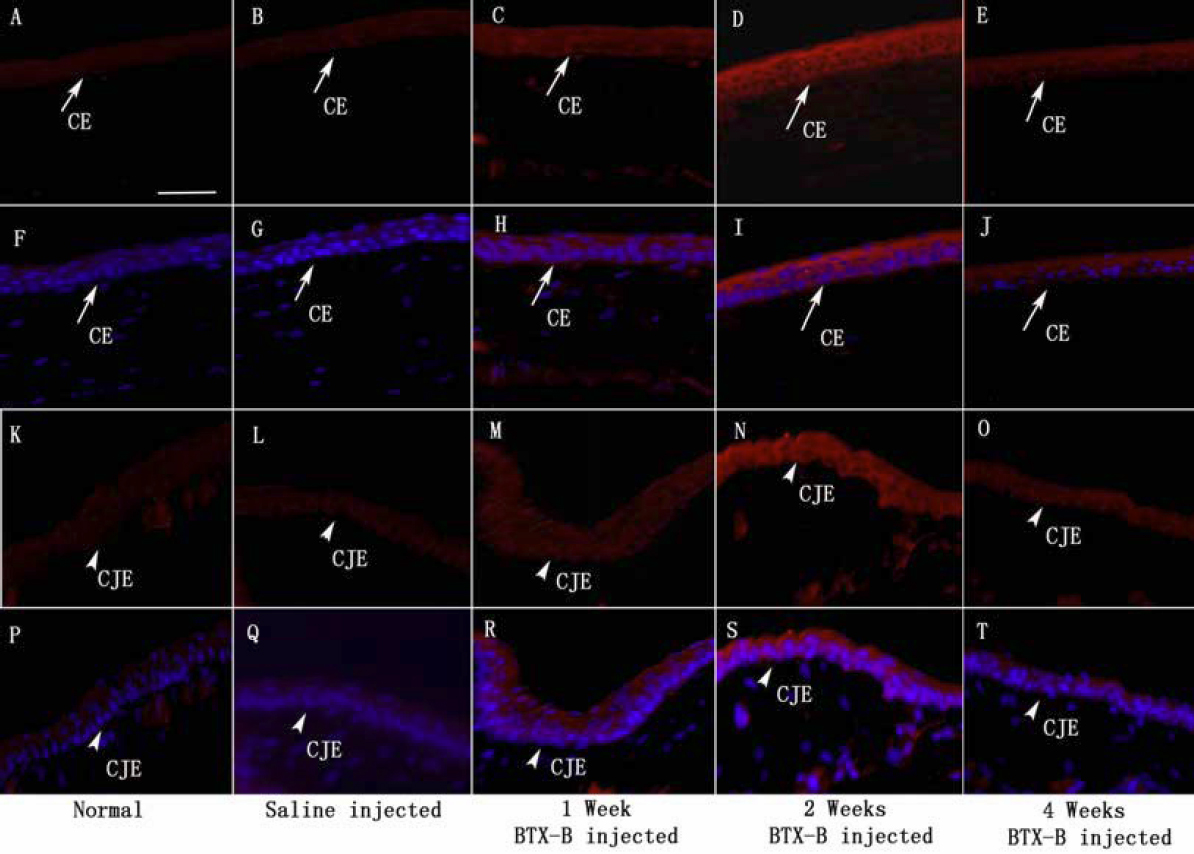
 Figure 3 of
Zhu, Mol Vis 2012; 18:1803-1812.
Figure 3 of
Zhu, Mol Vis 2012; 18:1803-1812.