Figure 5. Slit-lamp photographs and pedigree of severe family I. A, B: Patient 8, aged 35; C, D: Patient 9, aged 38; E, F: Patient 10, aged 59. A, C, E: Right eye, B, D, F: Left eye. Confluent granular deposits, prominent lattice deposits, and anterior haze occupying the visual axis was observed
in both eyes of all family members. They showed a more severe phenotype than other patients of a similar age and members within
the family showed a similar pattern. G: Pedigree of severe family I. Circles represent women, squares represent men. The filled symbols indicate affected individuals.
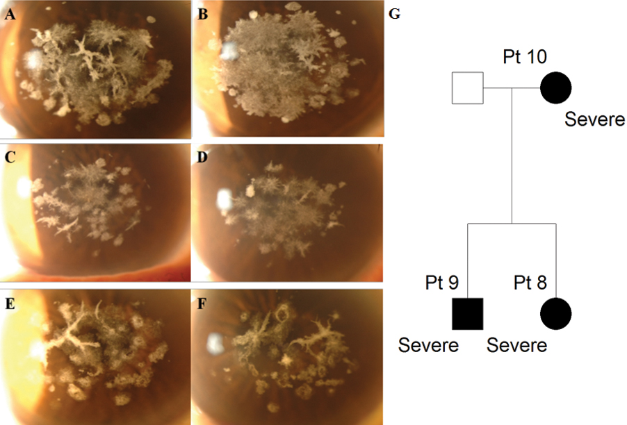
 Figure 5 of
Han, Mol Vis 2012; 18:1755-1762.
Figure 5 of
Han, Mol Vis 2012; 18:1755-1762.