Figure 4. Slit-lamp photographs and pedigree of mild family II. A, B: Patient 5, aged 33; C, D: Patient 6, aged 34; E, F: Patient 7, aged 62. A, C, E: Right eye, B, D, F: Left eye. A: Several granular and linear corneal opacities were observed. B: A few small granular deposits and linear opacities were observed. C, D: Opacities of round and linear shapes were observed centrally in the anterior stroma in both eyes. E: A small granular deposit was observed around the pupillary margin at 12 o’clock (black arrowhead). F: Faint granular opacities at the 1, 4, 5, and 7 o’clock positions were observed (black arrowheads). G: Pedigree of mild family II. Circles represent women, squares represent men. The filled symbols indicate affected individuals.
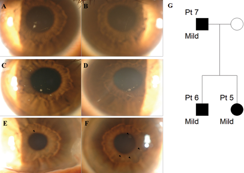
 Figure 4 of
Han, Mol Vis 2012; 18:1755-1762.
Figure 4 of
Han, Mol Vis 2012; 18:1755-1762.