Figure 2. Photographic demonstration of aniridia expression in family members. The eyes of patient II:1 exhibit iris hypoplasia and
lenses opacity (A), whereas those of patient III:1 had aniridia and transparent lenses (B). Pentacam photo shows the normal anterior segment picture of the patient II:1 from the family (C), and that of patient III:1 shows the same. The eyes of the family exhibited foveal hypoplasia by eyeground photography,
including II:1 and III:1 (D).
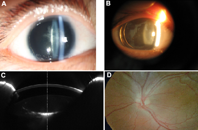
 Figure 2 of
Kang, Mol Vis 2012; 18:1750-1754.
Figure 2 of
Kang, Mol Vis 2012; 18:1750-1754.