Figure 7. The effect of Y-27632 on proteoglycans after superficial keratectomy. No differences in KS-GAG distribution were evident from
immunohistochemistry with the 5D4 antibody. Electron microscopy revealed that large cuprolinic blue-stained proteoglycan filaments,
typical of healing stromal scars and presumably of the chondroitin sulfate/ dermatan sulfate subfamily, were present in both
groups. Scale bar: top; 100 μm, below; 0.5 μm.
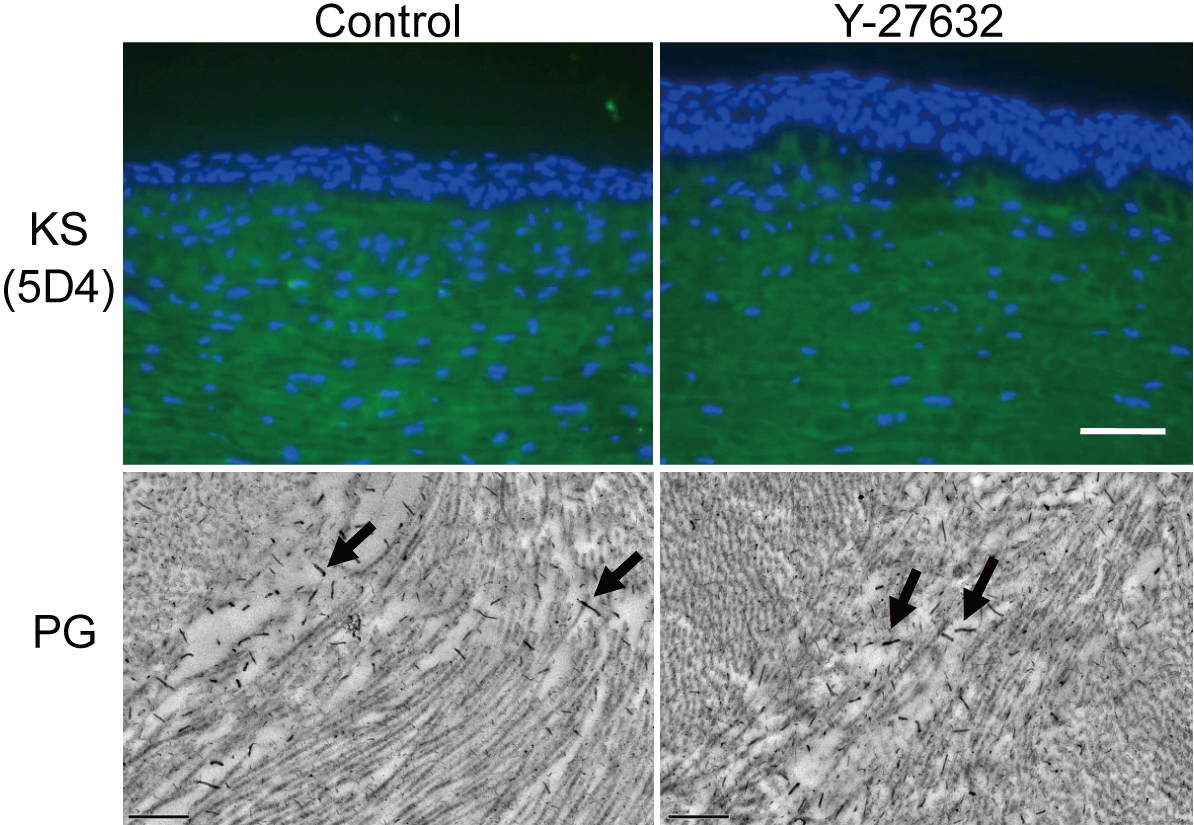
 Figure 7 of
Yamamoto, Mol Vis 2012; 18:1727-1739.
Figure 7 of
Yamamoto, Mol Vis 2012; 18:1727-1739.