Figure 1. Clinical findings in fundus
albipunctatus patients. A: Fundus photograph of the left
eye of patient MOL0427–1 at the age of 19 years showing multiple
small white-yellow retinal spots and flecks with relative
sparing of the fovea. B: ERG recordings of patient
MOL0427–1 under scotopic conditions showing markedly reduced
mixed cone-rod responses after 30 min of dark adaptation in both
eyes. Following 3 h of dark adaptation in the right eye, the
responses significantly improved and reached the normal range. C:
Fundus photograph of the left eye of patient MOL0091–2 at 29
years of age. Small patches of yellowish atrophy at the fovea
are apparent (arrowheads) in addition to preexisting punctata
(asterisks).
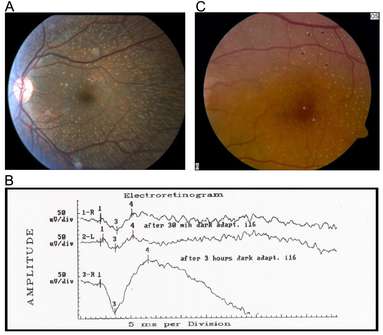
 Figure 1
of Pras, Mol Vis 2012; 18:1712-1718.
Figure 1
of Pras, Mol Vis 2012; 18:1712-1718.