Figure 5 of
Hazra, Mol Vis 2012; 18:1701-1711.
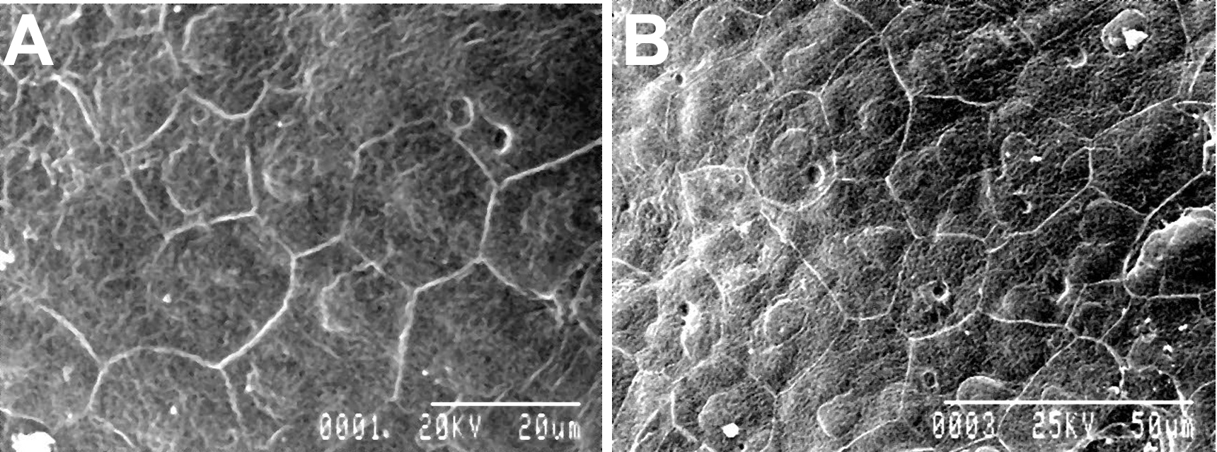
Figure 5.
Scanning electron microscopy shows well defined corneal endothelial cells with unaltered cellular junction in both control (
A
) and EDTA treated (
B
) eyes.
 Figure 5
of Hazra, Mol Vis 2012; 18:1701-1711.
Figure 5
of Hazra, Mol Vis 2012; 18:1701-1711.