Figure 3. Effect of EDTA on posterior
capsular opacification. Miyake Apple posterior view shows
opacification of posterior capsule in control eyes (A), a
clear posterior capsule in EDTA treated eyes (B); and
quantitative representation for degree of opacification (C).
Histological section of the lens capsule from control (D)
eyes shows increased number of LECs arranged in multilayers
whereas reduced cellular proliferation in EDTA-treated eyes (E);
and quantitative representation (F). Immunohistochemistry
shows increased expression of MMP2 in the lens capsule from
control eyes (G) and reduced expression in the lens
capsule from EDTA treated eyes (H), quantitative
representation (I), corresponding DAPI stained sections (J)
and (K). Magnification: 20×; p<0.05.
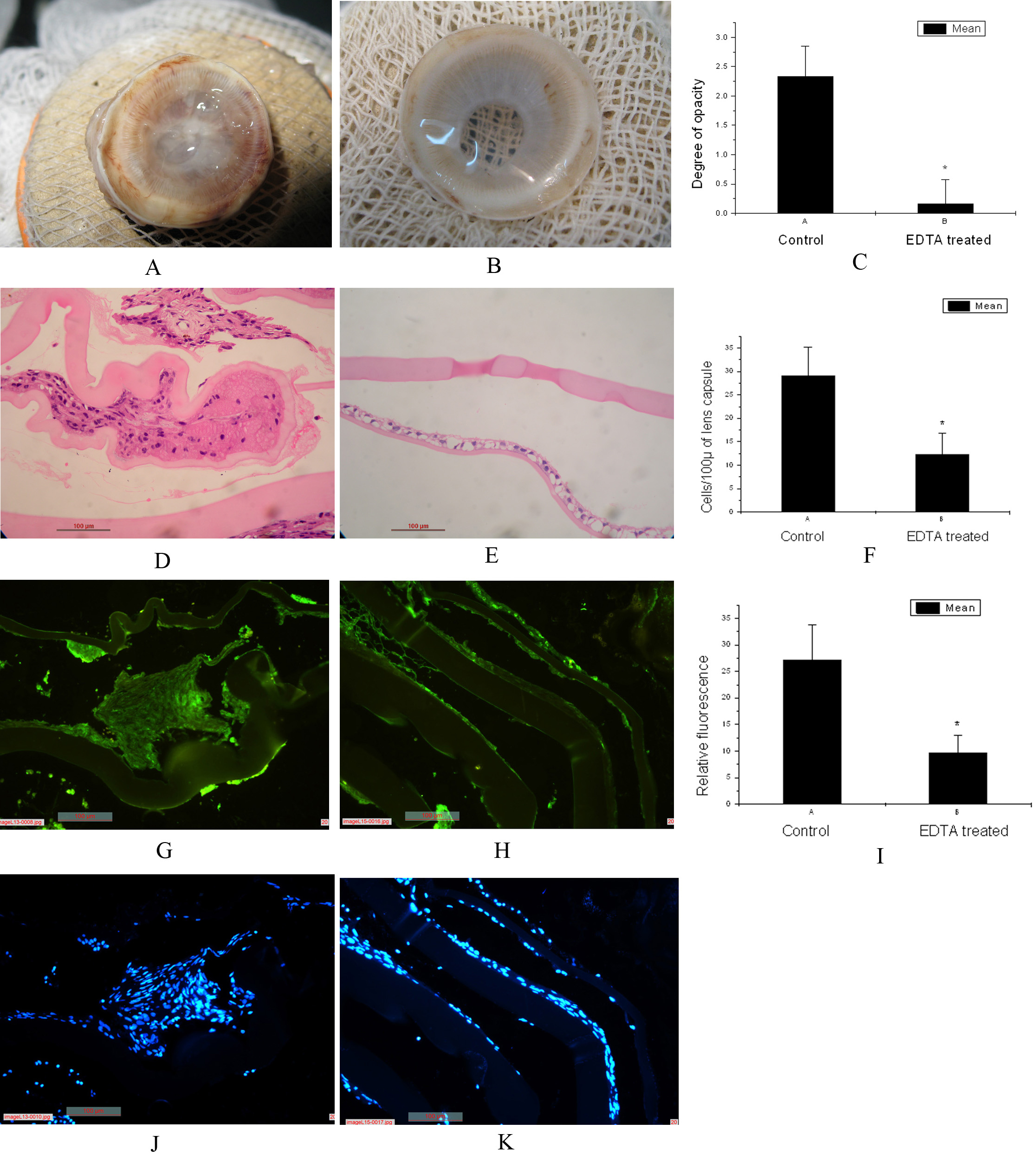
 Figure 3
of Hazra, Mol Vis 2012; 18:1701-1711.
Figure 3
of Hazra, Mol Vis 2012; 18:1701-1711.