Figure 5. We used Boyden chambers and
paxillin immunofluorescence staining assays to determine that
effects of heparanase expression levels on human retinal
vascular endothelial cell migration. A: The human
retinal vascular endothelial cells (HRECs) that are stained with
crystal violet are those that migrated across the extracellular
matrix (ECM) in the specialized migration chamber by migrating
through the polycarbonate membrane to the lower surface of the
membrane (original magnification 200×). B: Paxillin
immunofluorescence staining indicated positive paxillin protein
expression in every group. HPSE-LV significantly inhibited HREC
paxillin protein expression compared with the Con-LV-Glu30 and
Glu30 groups. The paxillin protein expression in the Glu30 group
was significantly higher than in the HPSE-LV-Glu30 and Glu5
groups (the yellow arrow indicates paxillin). C:
Heparanase small interfering RNA recombinant lentiviral vector
(HPSE-LV) significantly decreased the HRECs’ migration ability.
Bars indicate the mean±SD (p<0.05). The HPSE-LV-Glu30 group
showed a markedly decreased migration ability compared to the
Con-LV-Glu30 (p<0.01) and Glu30 groups, which migrated
significantly more than the HPSE-LV-Glu30 and Glu5 groups
(●p<0.01 versus Glu5; ▲p<0.01 versus HPSE-LV-Glu30). Each
group experiment was repeated 3 times (n=3).
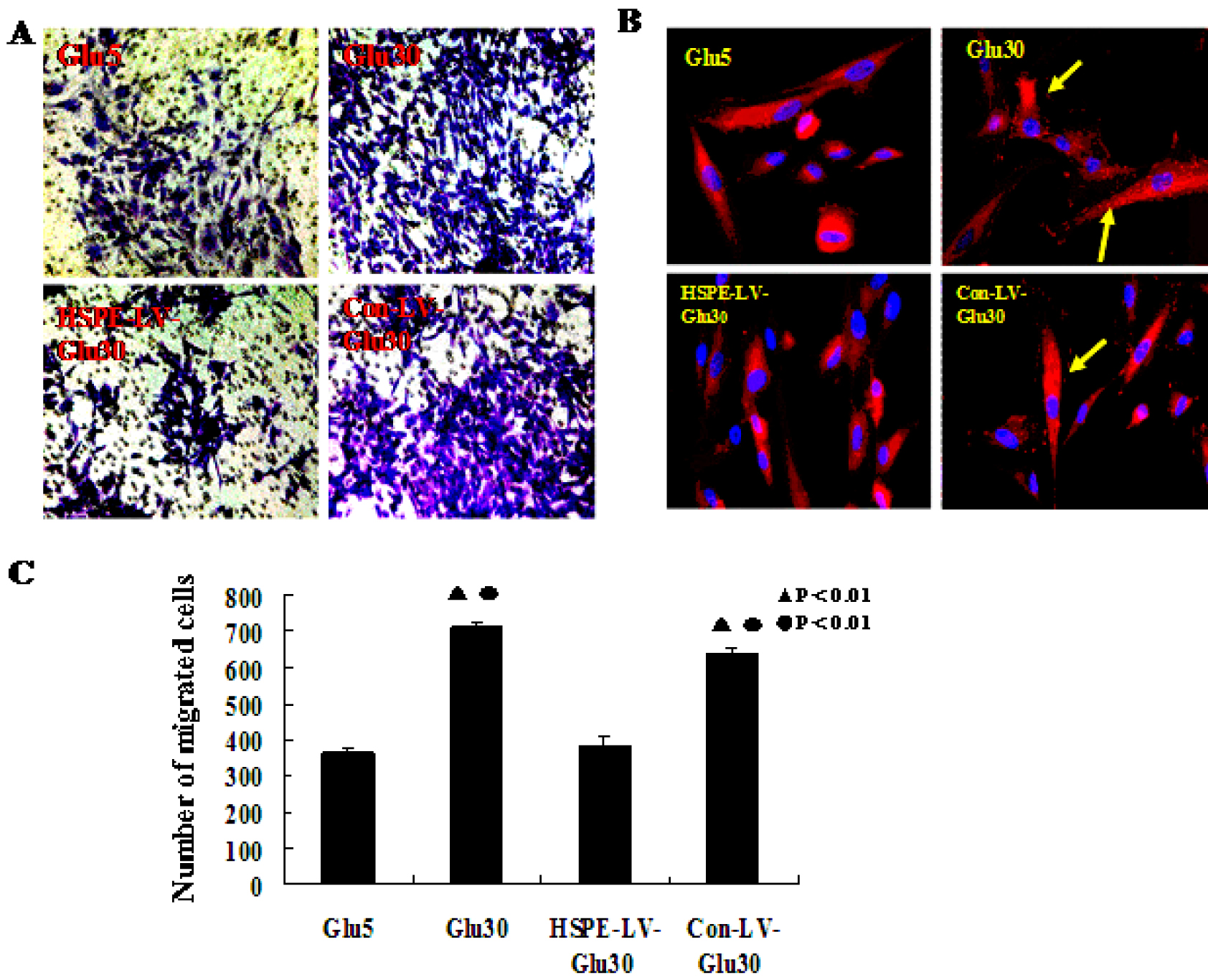
 Figure 5
of Yuan, Mol Vis 2012; 18:1684-1695.
Figure 5
of Yuan, Mol Vis 2012; 18:1684-1695.