Figure 1. Heparanase expression in human retinal vascular endothelial cells under high-glucose conditions for 48 h is increased than
in the Glu5 and mannitol control groups. A, B: We used western blotting to determine that the expression of heparanase in human retinal vascular endothelial cells (HRECs)
grown in conditioned medium. The band that represents heparanase expression was more intense in the Glu30 group than in the
Glu5 and mannitol control groups. Each group experiment was repeated 3 times (n=3). * compared with the Glu30 group (p<0.01);
** compared with the Glu5 and mannitol control groups (p<0.01).
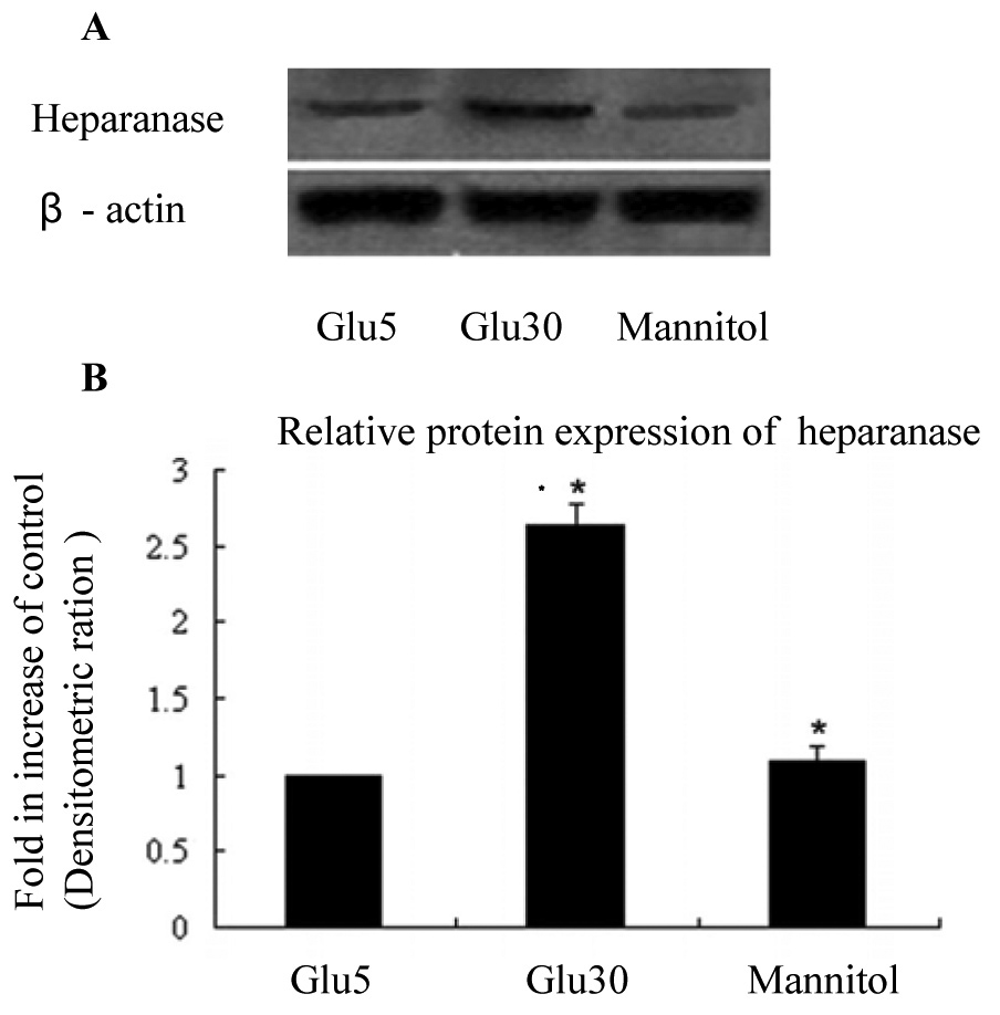
 Figure 1 of
Yuan, Mol Vis 2012; 18:1684-1695.
Figure 1 of
Yuan, Mol Vis 2012; 18:1684-1695.