Figure 8. Ultrastructure of optic
nerve axonal loss. A: One year after injection of scAAV-GFP,
transmission electron micrographs disclosed optic nerve axons
(a) of various sizes enveloped by myelin sheaths. Astroglial
cell processes (g) coursed between fibers of the optic nerve. B:
In this same animal, the opposite eye injected with the MTS
scAAV mutant ND4 had a marked decrease in axonal
density. Many empty spaces (e) were present where axons were
apparently lost in these atrophic optic nerves and degenerating
axonal profiles were evident (d). C: In a different
animal, the optic nerve of the control eye injected with scAAV-GFP
had normal axons (a) with the only empty space the lumen of a
blood vessel (L). D: In this animal, the opposite eye
injected with the MTS scAAV mutant ND4 had a marked decrease in
axonal density. Many empty spaces (e) were present where axons
were lost. a=optic nerve axon, e=empty space, g=astroglial
process, d=degenerating axon, L=lumen.
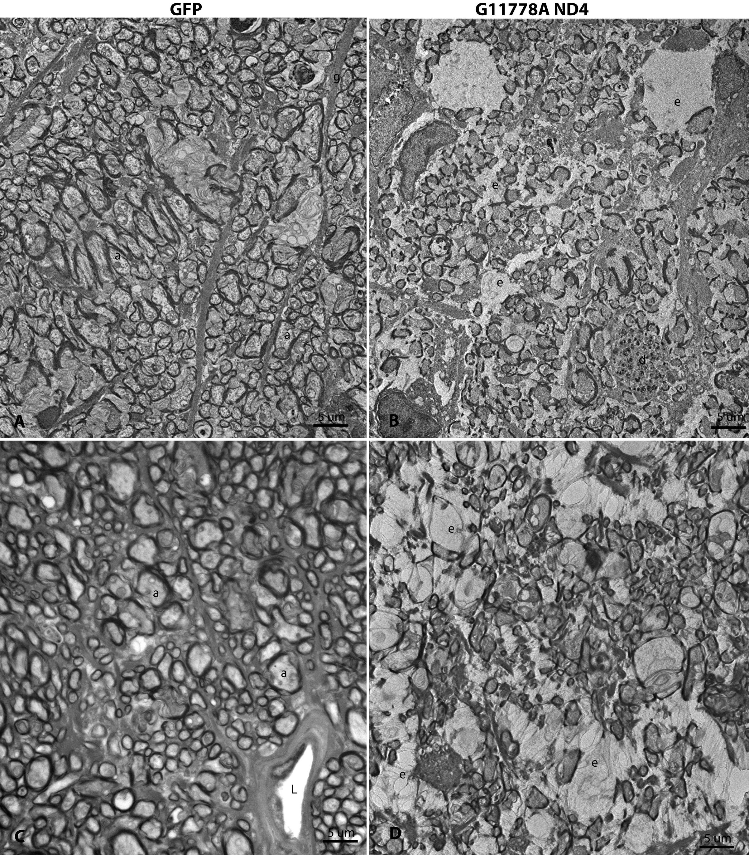
 Figure 8
of Yu, Mol Vis 2012; 18:1668-1683.
Figure 8
of Yu, Mol Vis 2012; 18:1668-1683.