Figure 5. SD-OCT imaging of optic
disc edema and optic atrophy. A: Spectral domain optical
coherence tomography (SD-OCT) of right eyes injected with the
mutant human ND4 packaged with the MTS scAAV revealed
swelling of the optic nerve head (arrows) commencing at 1 month
post injection. A focal increase in the thickness of the RGC and
the inner plexiform layer (IPL) is apparent just to the right of
the swollen optic nerve head. B: The control eye
injected with scAAV-GFP showed the normal anatomy of the mouse
optic nerve head. C: One year post injection, optic
nerve head atrophy was apparent in the mutant ND4 injected eyes.
D: The contralateral GFP injected control eyes maintained
normal optic nerve head anatomy. E: One year post
injection, focal thinning with loss of the inner retina was also
apparent in an experimental eye, but this finding was not seen
in any of the control eyes (F). RGC=retinal ganglion cell
layer; IPL=inner plexiform layer.
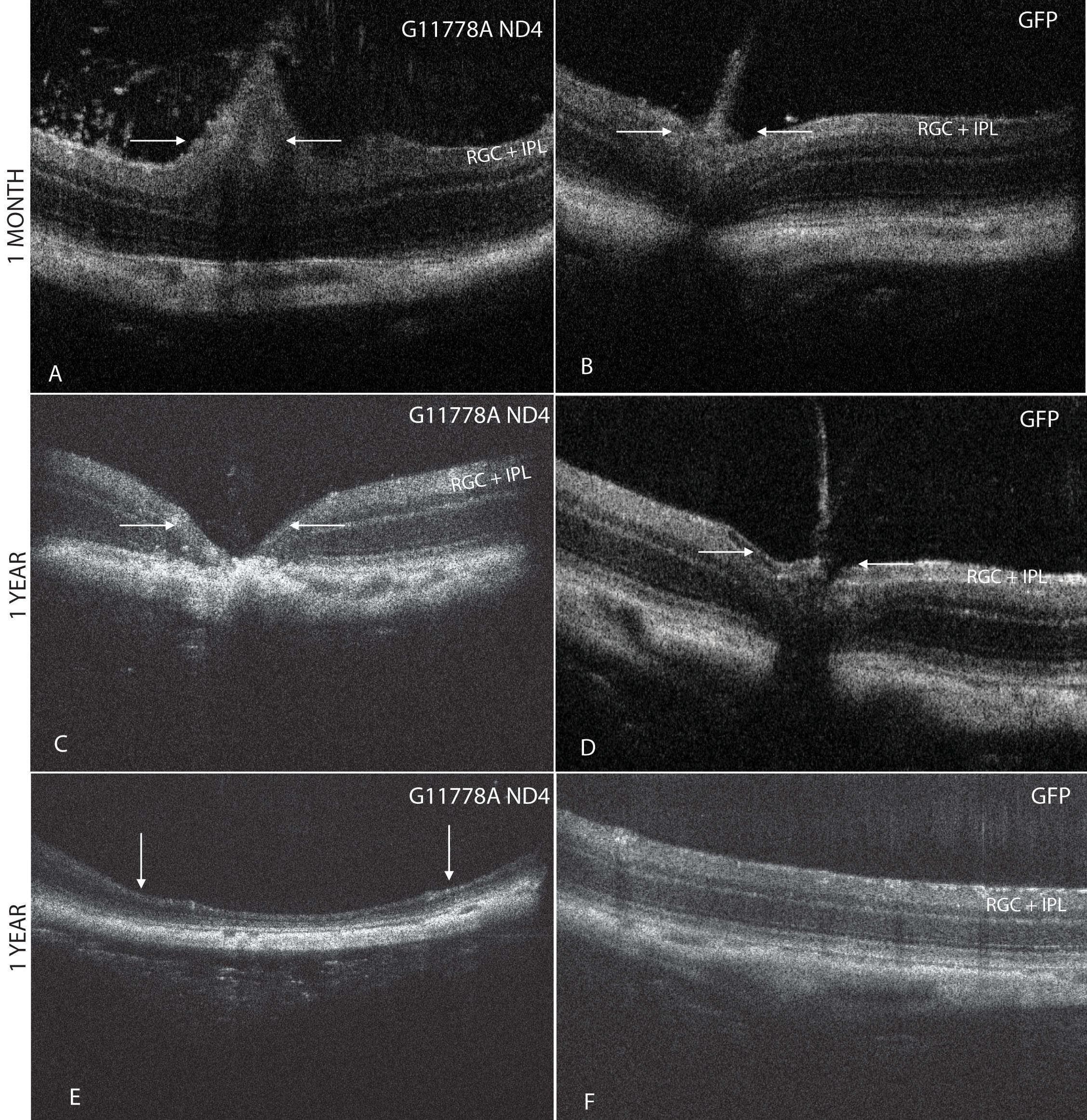
 Figure 5
of Yu, Mol Vis 2012; 18:1668-1683.
Figure 5
of Yu, Mol Vis 2012; 18:1668-1683.