Figure 3. Wild-type human ND4 expression in RGCs does not cause visual loss in mice. Confocal immunofluorescence micrographs show DAPI
stained nuclear layers of the murine retina infected 7 weeks earlier with MTS scAAV containing wild-type normal human ND4FLAG (A). The RGC layer and dendrites extending into the inner nuclear layer (INL) are labeled with an antibody against Thy1.2. The
outer nuclear layer (ONL) is not stained by Thy1.2 (B). RGCs expressing human ND4FLAG are labeled by an anti-FLAG antibody (C) that colocalizes to Thy1.2 and has a perinuclear distribution characteristic of mitochondria surrounding the DAPI-stained
RGC nuclei (D). A scatterplot (E) and a plot of representative PERG waveform (F) show that 7 weeks after AAV injections there were no differences in amplitude between the eyes injected with wild-type human
mitochondrial (m) ND4 relative to the eyes that received no ocular gene injections.
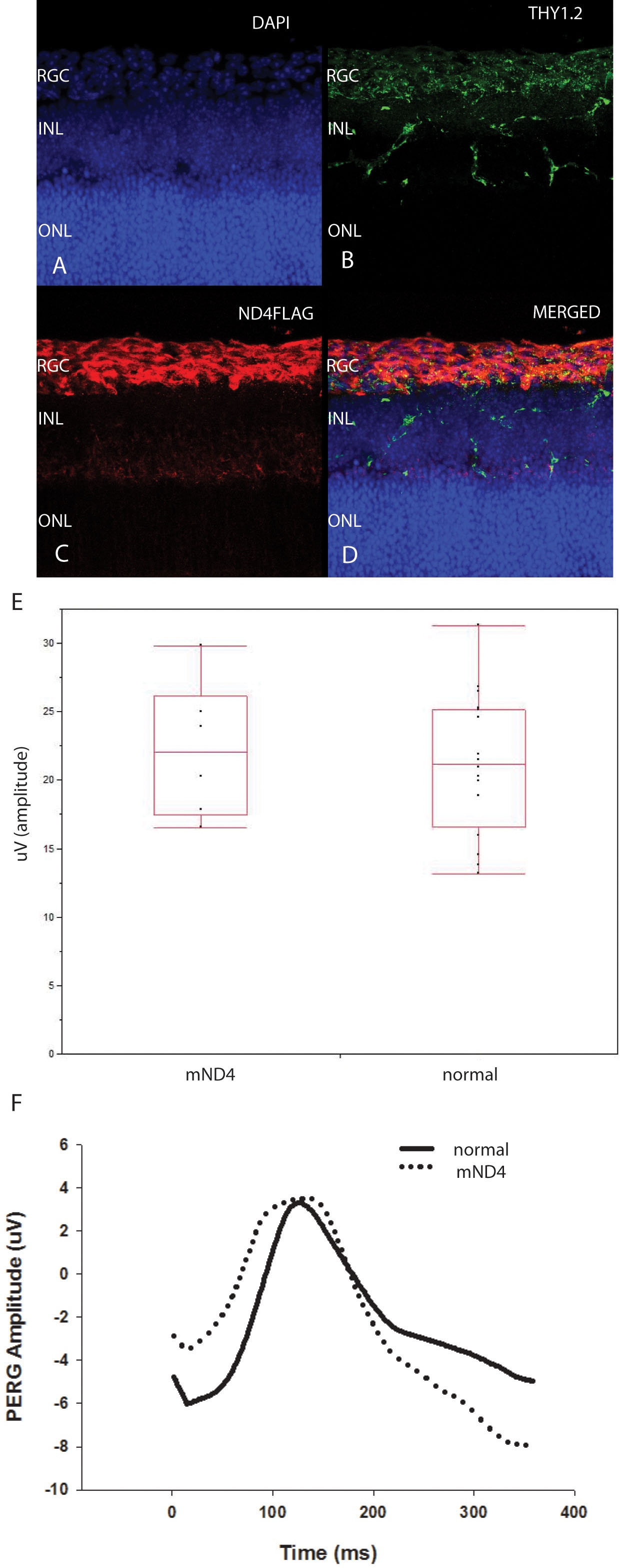
 Figure 3 of
Yu, Mol Vis 2012; 18:1668-1683.
Figure 3 of
Yu, Mol Vis 2012; 18:1668-1683.