Figure 1. MTS AAV-capsid localizes
within mitochondria where the delivered wild-type human ND4
is translated in cultured cells. A: Illustration of the
COX8 MTS fused in frame with GFP and inserted into the
VP2 capsid of AAV. B: Illustration of the mitochondrial
heavy strand promoter (HSP) driving expression of the
mitochondrial mutant human ND4 subunit of complex I to which is
appended a FLAG epitope. C: Fluorescence microscopy of
live cultured cells infected with the COX8GFP VP2 MTS
AAV revealed punctate and perinuclear expression of GFP
suggestive of mitochondrial localization. D: MitoTracker
Green staining of mitochondria surrounds nuclei labeled with
DAPI. E: Transmission electron microscopy shows silver
enhanced GFP immunogold inside mitochondria as well as within
the nucleus. F: ND4FLAG immunogold is evident only
within the mitochondria. Abbreviations m=mitochondria and
n=nucleus.
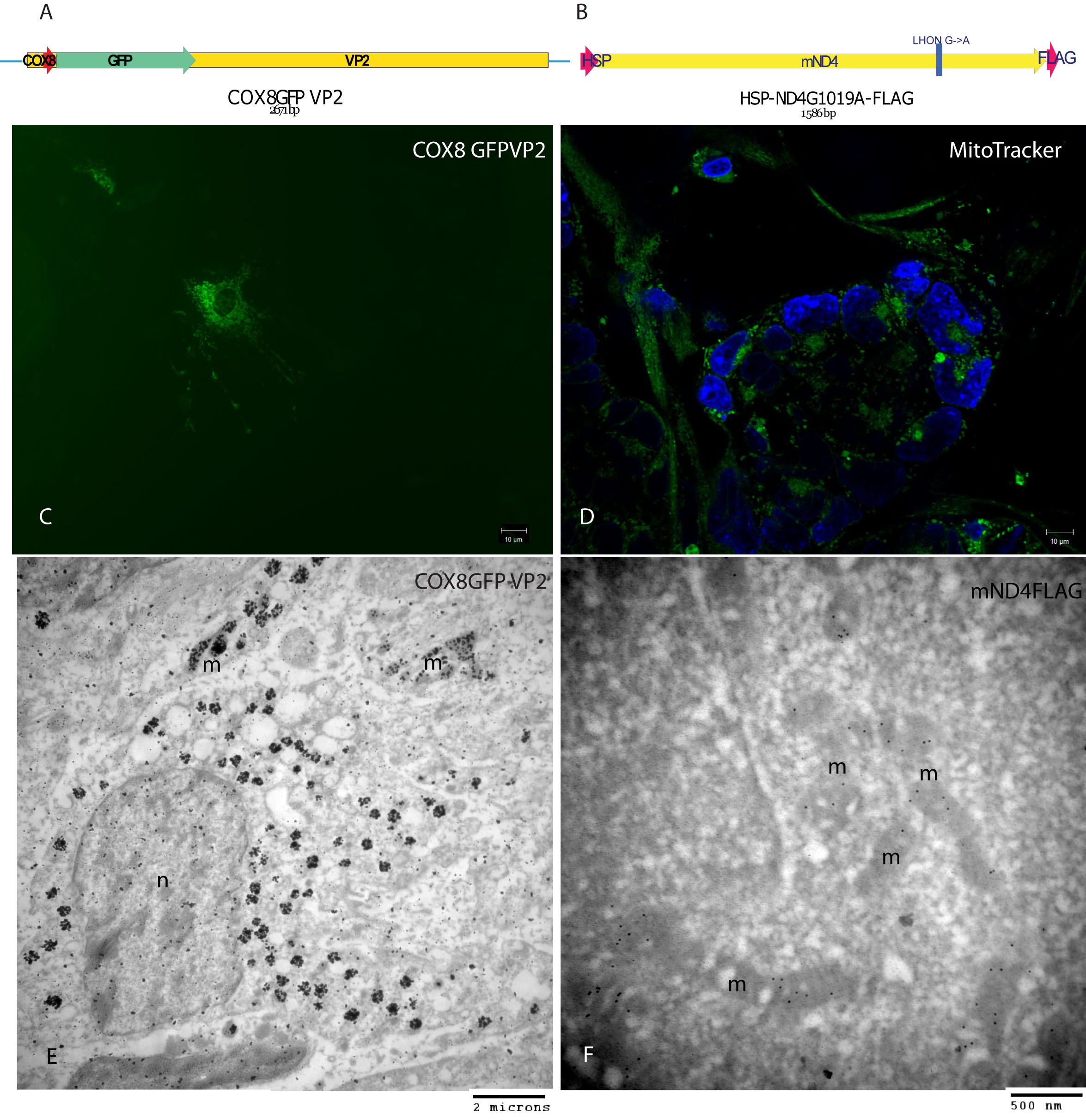
 Figure 1
of Yu, Mol Vis 2012; 18:1668-1683.
Figure 1
of Yu, Mol Vis 2012; 18:1668-1683.