Figure 2. The effects of
Immunohistochemistry staining for retina sections were accessed
in this study. A-C: The retina sections stained
for heparanase positive cells were show. A: It shows a
normal mouse retina section on postnatal day 17 (P17). B:
It displays the retina section in oxygen-induced retinopathy
(OIR) mice. C: It shows PI-88 treatment of an OIR mouse
retina. In all of the sections, the red arrow points to
heparanase positive cells. D-F: The retina
sections stained for vascular endothelial growth factor (VEGF)
positive cells were show. D: It shows a normal mouse
retina section on P17. E: It displays the retina section
in OIR mice; F: It shows phosphomannopentaose sulfate (PI-88)
treatment of an OIR mice retina. In all of the sections, the red
arrow points to VEGF positive cells. G: The percentages
of heparanase-positive cells in retina tissue by
immunohistochemistry were as follows: The percentage of Group A
was 32.00±7.00; the percentage of Group B was 51.33±1.53; the
percentage Group C was 32.00±3.61. H: The percentages of
VEGF-positive cells in retina tissue by immunohistochemistry
were as follows: The percentage of Group A was 65.00±2.00. The
percentage of Group B was 101.33±10.11; The percentage Group C
was 19.00±2.65. The differences among groups were significant
(p<0.0001).
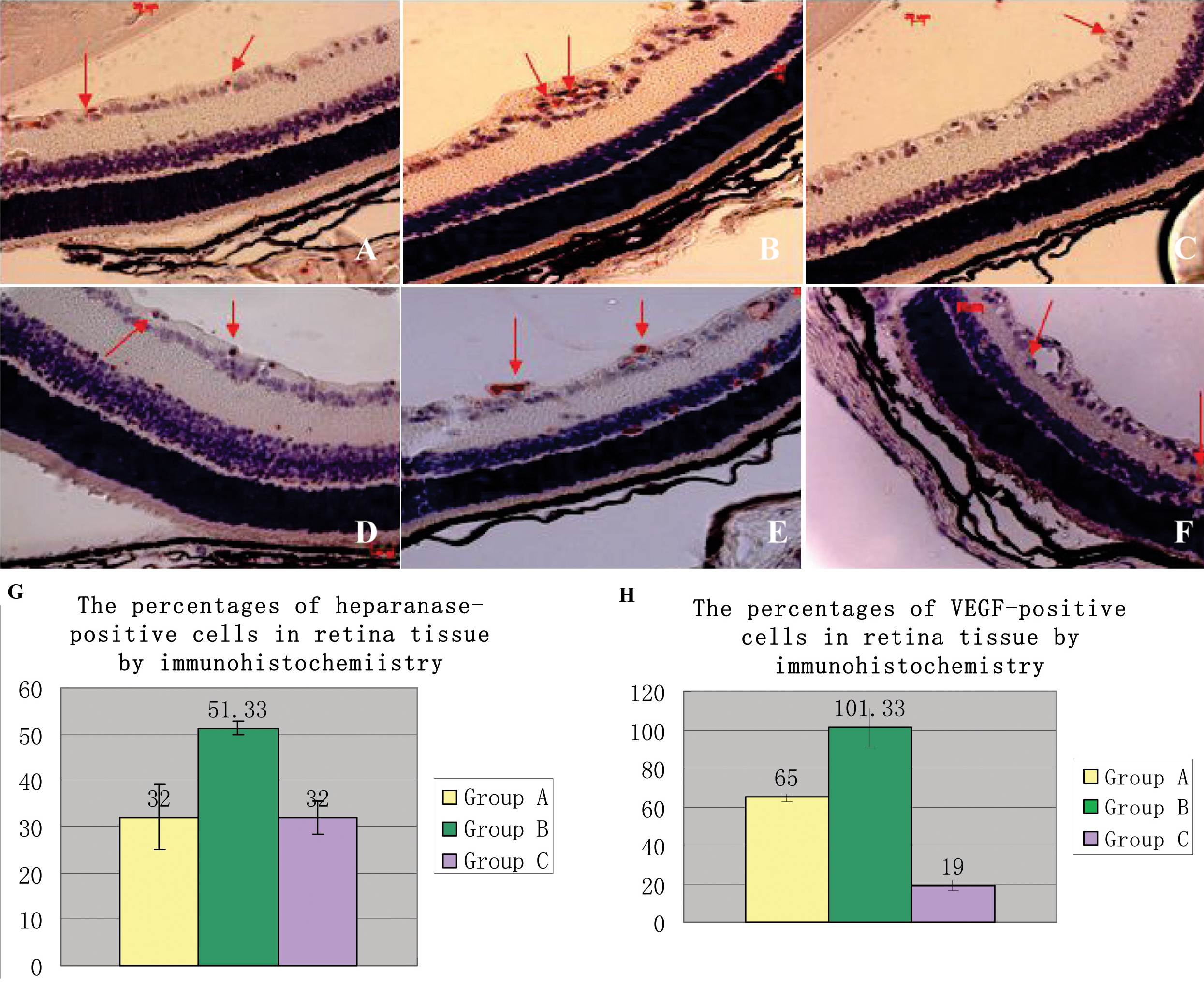
 Figure 2
of Liang, Mol Vis 2012; 18:1649-1657.
Figure 2
of Liang, Mol Vis 2012; 18:1649-1657.