Figure 1. Fluorescein isothiocyanate-dextran (FITC) perfusion of the retinal blood vessels was observed under a fluorescent microscope
in postnatal day 17 (P17) oxygen-induced retinopathy (OIR) retinas. A: Mice were perfused with FITC-labeled fluorescein sodium in normal oxygen pressure. B: Mice were perfused with FITC-labeled fluorescein sodium in hypoxia. Fluorescence images showing retinal vessels were tortuous
and expanded in volume. Capillary hemangioma was observed, and retinal vascular morphology exhibited abnormal distribution
at the junction of the perfused area and non-infused area. Significant expansion of large vessels, large avascular area, and
extensive angiogenesis were observed. There are non-vascular area and neovascular tuffs in oxygen-induced retinopathy (OIR)
mice (arrow).
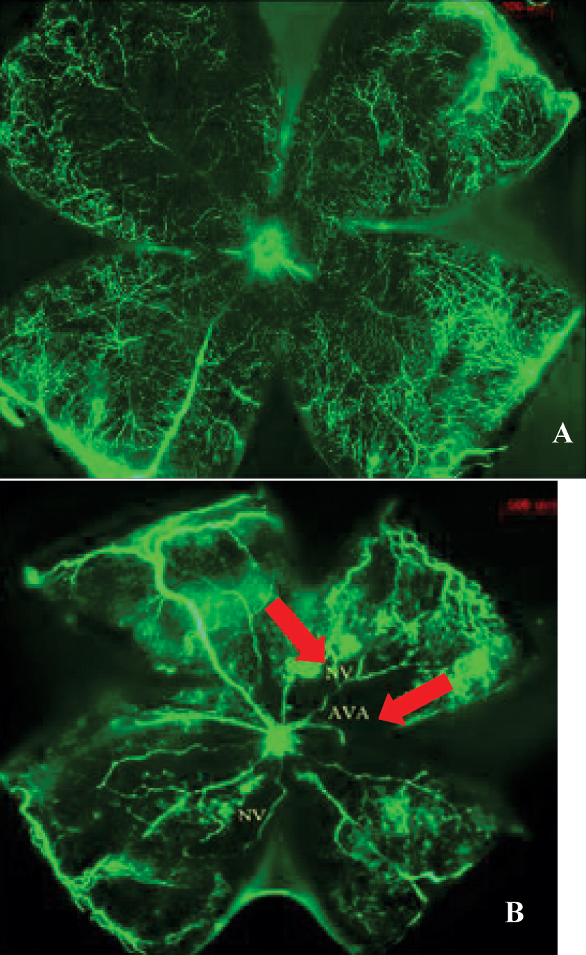
 Figure 1 of
Liang, Mol Vis 2012; 18:1649-1657.
Figure 1 of
Liang, Mol Vis 2012; 18:1649-1657.