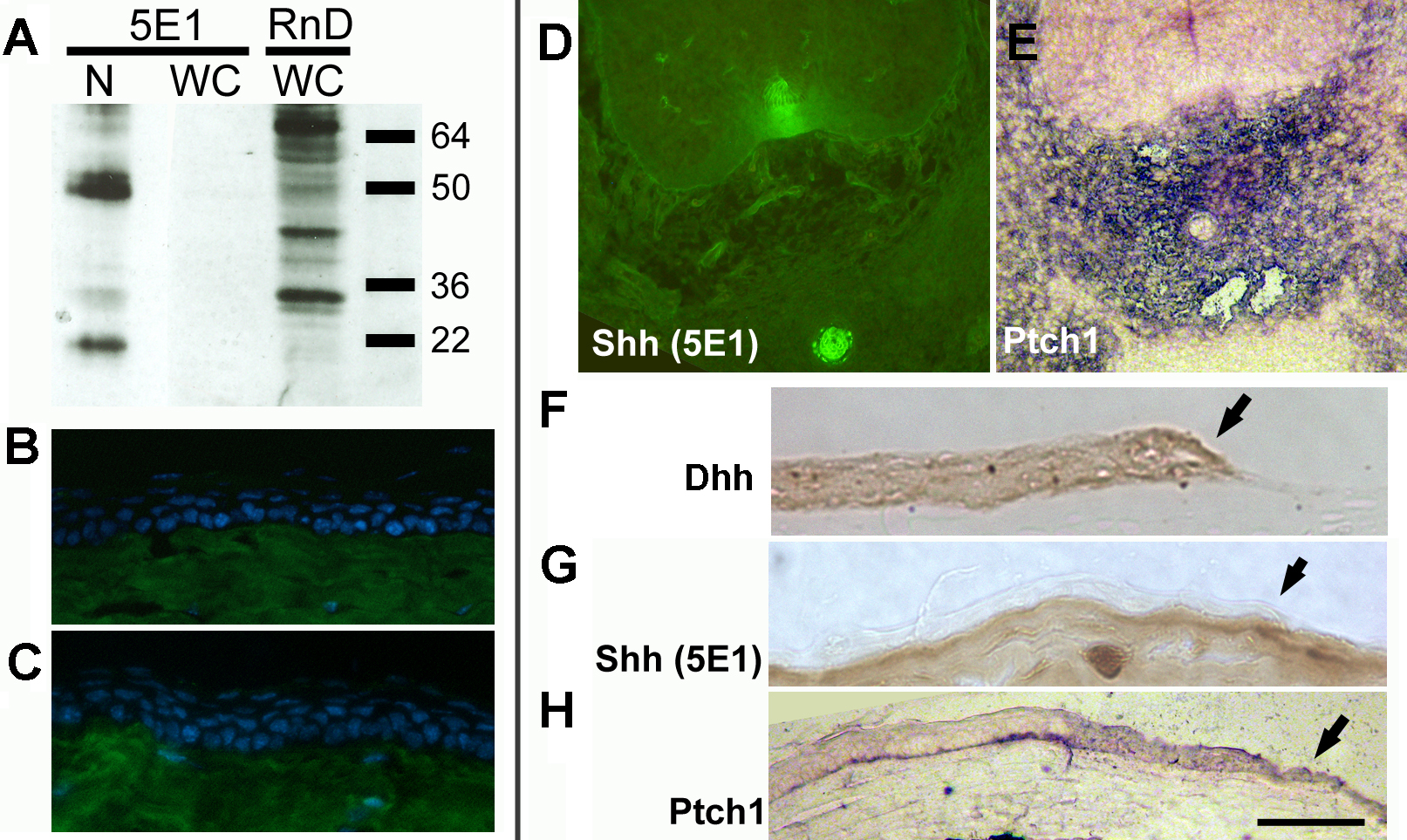
Figure 5. Expression of Hedgehog
signaling pathway components in the adult corneal epithelium.
A:
western blot analysis of embryonic notochord/neural tissue (N)
or wounded adult corneas using the 5E1 mouse anti-Shh-N
monoclonal (
Developmental
Studies Hybridoma Bank) or R&D Systems goat
anti-mShh-N polycolonal AF464. 5E1 recognizes 2 major bands at
sizes predicted for Shh isoforms in embryonic tissue (left hand
lane), but detects no Shh-N in the wounded adult cornea (middle
lane). The goat polyclonal recognizes multiple non-Shh bands in
wounded corneas.
B,
C: No Shh is detected by
immunohistochemistry in the wild-type adult corneal epithelium
by the 5E1 antibody (
B), although there is strong
background staining of the underlying corneal stroma by the
Alexa-488 conjugated goat anti-mouse IgG
1 secondary
antibody (A21121; Molecular Probes), as shown in
C the
negative (no primary antibody) control.
D: Shh
localization in transverse section of E12.5 notochord and floor
plate (immunohistochemistry using 5E1 monoclonal antibody) and
EPtch1
expression (purple) upregulated in tissues around the notochord
shown by in situ hybridization. The sense control was blank
(data not shown).
F: Localization of Dhh by
immunohistochemistry with DAB (brown) endpoint in the wounded
corneal epithelium. Wound edge is indicated by an arrow.
G:
No Shh in the corneal epithelium at a healing wound (arrow) edge
shown by immunohistochemistry using the 5E1 monoclonal antibody.
H: Upregulation of
Ptch1 at the wound edge
(arrow) is evidence that Hedgehog signaling is active. Scale
bar: 100 μm.
 Figure 5
of Kucerova, Mol Vis 2012; 18:139-150.
Figure 5
of Kucerova, Mol Vis 2012; 18:139-150.