Figure 5. Melatonin dose-dependently protected retinal pigment epithelial cells against H2O2 damage as tested by microculture tetrazoline test, compared with cells not treated with H2O2. Retinal pigment epithelial (RPE) cells were pretreated with melatonin (M) at concentrations of 10−10 M (M-10), 10−8 M (M-8), 10−6 M (M-6), and 10−4 M (M-4). After 24 h, 0.5 mM H2O2 (H) was added and cultured for 24 h. Cells not treated with H2O2 were used as negative controls (0). Cell viability was evaluated by the microculture tetrazoline test and expressed as percentages
of negative controls (mean±standard deviation [SD] in triplicate tests). Error bars represent SD A: Cell viability of cell treated with melatonin and H2O2 still significantly lower than that in cells cultured without H2O2 in the ARPE-19 cells (an immortal RPE cell line from a 19-year-old donor). B: The same was true in the primary-culture RPE cells. * p<0.05, compared with the negative controls (cells treated without
H2O2).
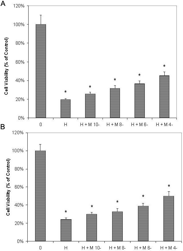
 Figure 5 of
Rosen, Mol Vis 2012; 18:1640-1648.
Figure 5 of
Rosen, Mol Vis 2012; 18:1640-1648.