Figure 3. The ARPE19 cells (an immortal retinal pigment epithelial cell line from a 19-year-old donor) were treated with melatonin (M)
at different concentrations. After 1 h, 24 h, and 48 h culture, 0.5 mM H2O2 (H) was added and cultured for 24 h. Cells cultured with H2O2 alone were used as the controls. Cell viability was evaluated by the microculture tetrazoline test and expressed as percentages
of controls (mean±SD in triplicate tests). Error bars represent SD A: Pretreatment with low concentrations of melatonin at 10−10 M (M-10) for 1 h, 24 h, and 48 h significantly protected cells against H2O2. B: Pretreatment with high concentrations of melatonin at 10−6 M (M-6) obtained similar results. The difference between cells cultured with and without melatonin at both high and low concentrations
was statistically significant at all three different pretreatment periods. *p<0.05, compared with the controls.
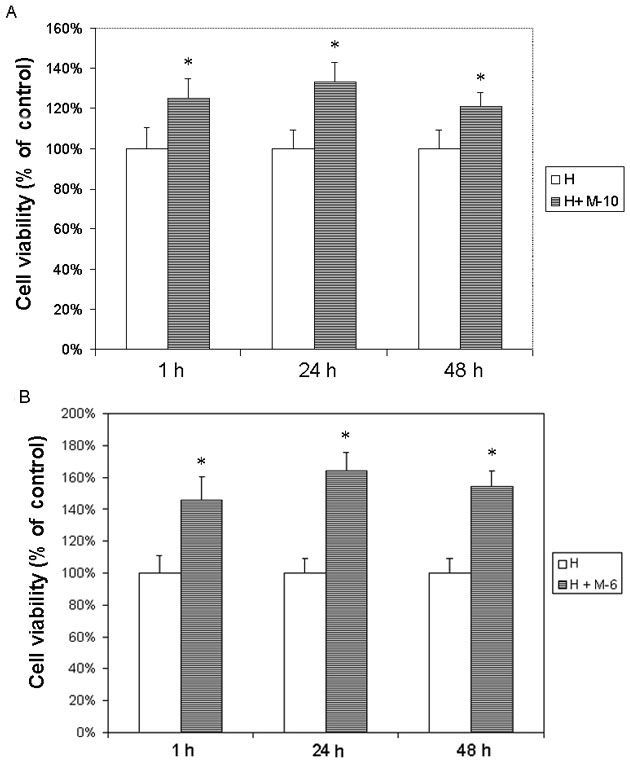
 Figure 3 of
Rosen, Mol Vis 2012; 18:1640-1648.
Figure 3 of
Rosen, Mol Vis 2012; 18:1640-1648.