Figure 6. YC-1 reduced RGC-5 cell
proliferation. A: YC-1 decreased the number of nuclei
and the Ki-67 cell proliferation marker expression in
RGC-5 cells using Cellomics® high-content screening
and the Columbus™ system. RGC-5 cells were treated with 5, 10,
20, and 40 µM YC-1 or 0.066% DMSO for 24 h. After fixation and
immunofluorescence staining, cells were detected and analyzed by
an HCS reader. Photos show fluorescence staining results of
Hoechst 33342 (blue) and Ki-67 (green). B:
The quantification results of HCS. * Indicates p<0.05
compared to the Hoechst-stained nuclei of the DMSO vehicle
group. # Indicates p<0.05 compared to the Ki-67-stained
cells of the DMSO vehicle group.
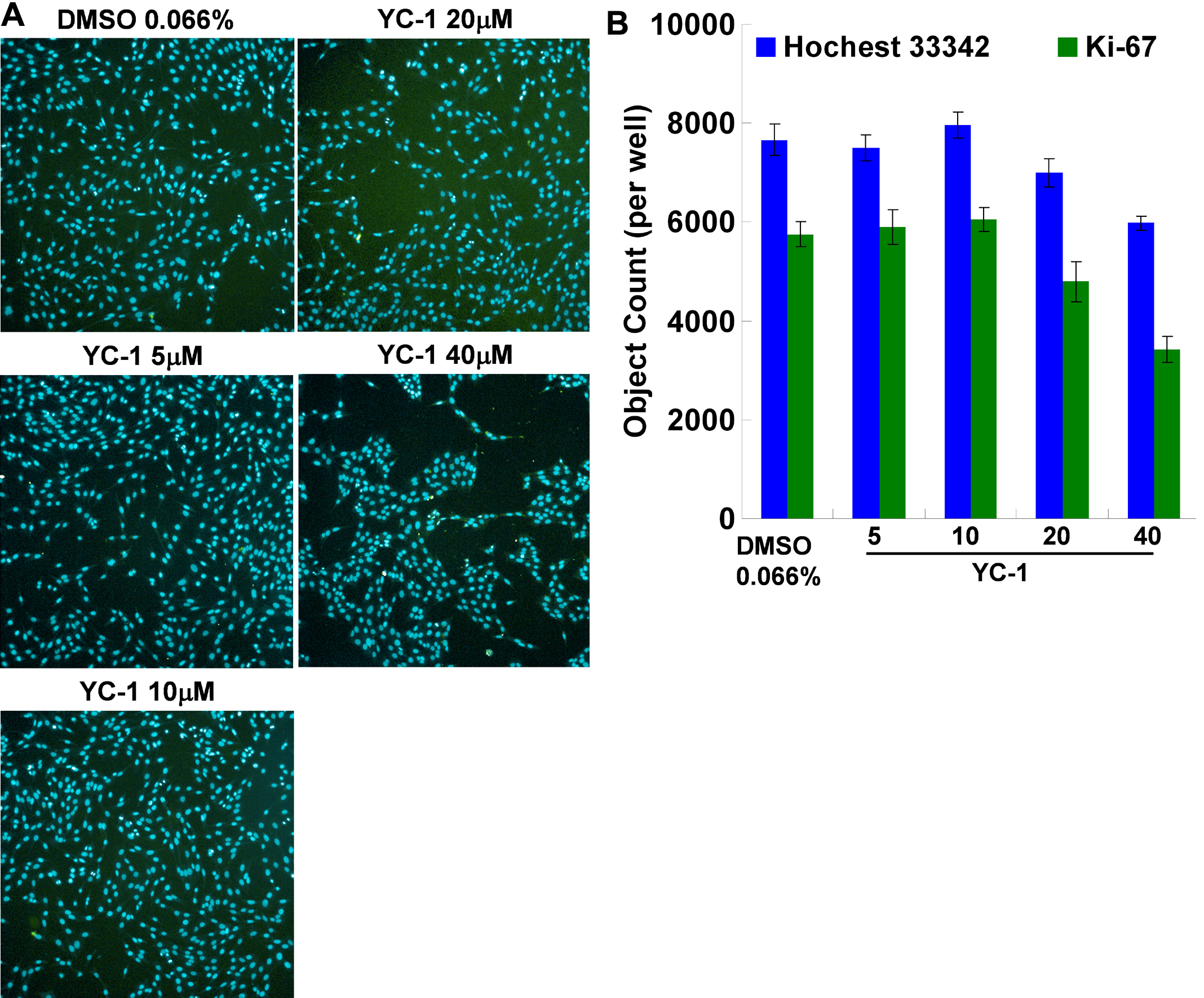
 Figure 6
of Tsui, Mol Vis 2012; 18:1594-1603.
Figure 6
of Tsui, Mol Vis 2012; 18:1594-1603.