Figure 3. An increase of the YC-1
exposure time resulted in a decrease of the RGC-5 cell density.
A: RGC-5 cells were exposed to 20 µM YC-1 for 4, 12, and
24 h. Morphological changes in cell density were observed with
light microscopy. B: Cell viability was detected by an
MTT assay of RGC-5 cells treated with YC-1 for different
incubation times. * Indicates p<0.05 compared to the DMSO
vehicle group.
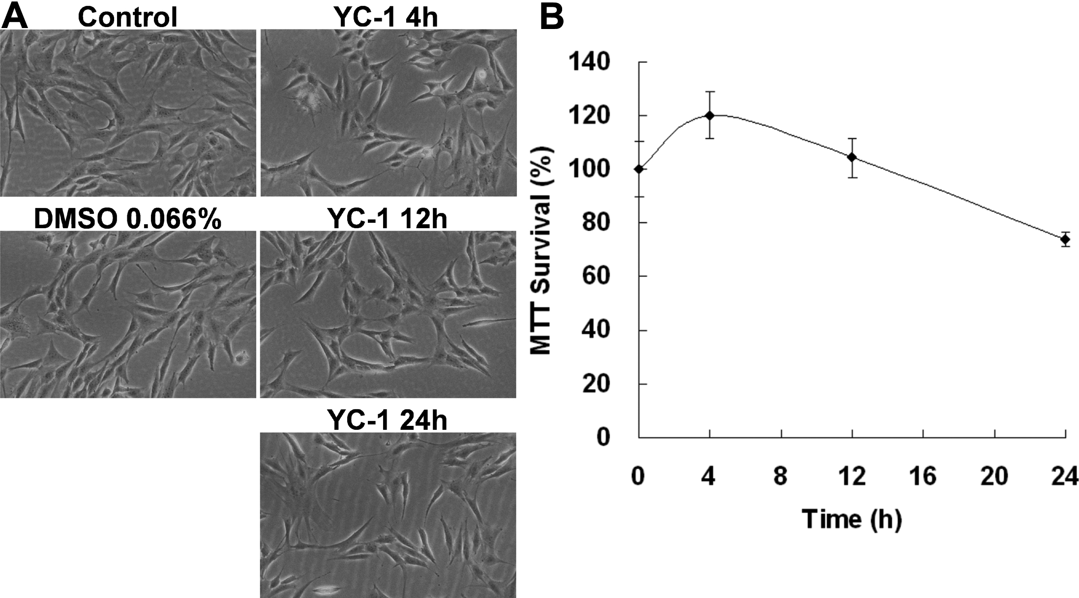
 Figure 3
of Tsui, Mol Vis 2012; 18:1594-1603.
Figure 3
of Tsui, Mol Vis 2012; 18:1594-1603.