Figure 2. YC-1 induced dose-dependent
retinal ganglion cell (RGC) cell death. A: RGC-5 cells
were exposed to 5, 10, 20, and 40 µM YC-1 and 0.066% DMSO for 24
h. Morphological changes in cell density were observed with
light microscopy. B: Cell viability was detected with an
MTT assay of increasing the YC-1 concentration to treat RGC-5
cells. * Indicates p<0.05 compared to the DMSO vehicle group.
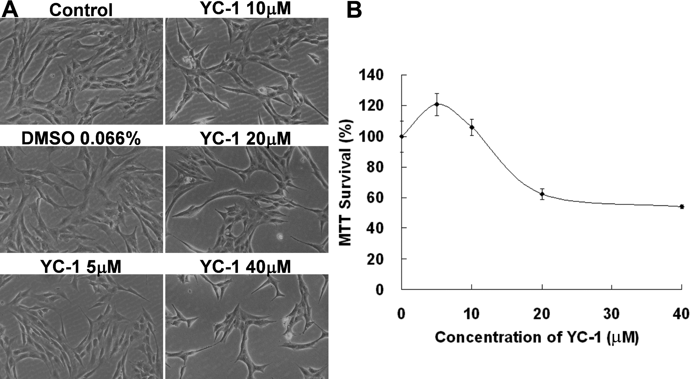
 Figure 2
of Tsui, Mol Vis 2012; 18:1594-1603.
Figure 2
of Tsui, Mol Vis 2012; 18:1594-1603.