Figure 5. Immunohistochemistry for
ANXA1 in ARPE-19 cells. ANXA1 immunostaining was detected in the
nucleus and cytoplasm of ARPE-19 cells (A-E).
After 60 and 120 min of infection, an intense immunoreactivity
of ANXA1 was detected in the infected cells (B and C)
as compared to control cells (A). In the later time
points of infection (240 min and 24 h), decreased expression of
ANXA1 was noted (D and E). Absence of ANXA1
immunostaining in ARPE-19 cells incubated with pre-immune serum
(F). Counterstain: Hematoxylin. Bars: 10 µm. G: Densitometric analysis of
ANXA1. Values (arbitrary units) are expressed as the mean±SEM of
sections analyzed from 10 cells/ group. *p<0.001 versus the
control group; #p<0.001 versus 60 min; +p<0.001
versus 120 min.
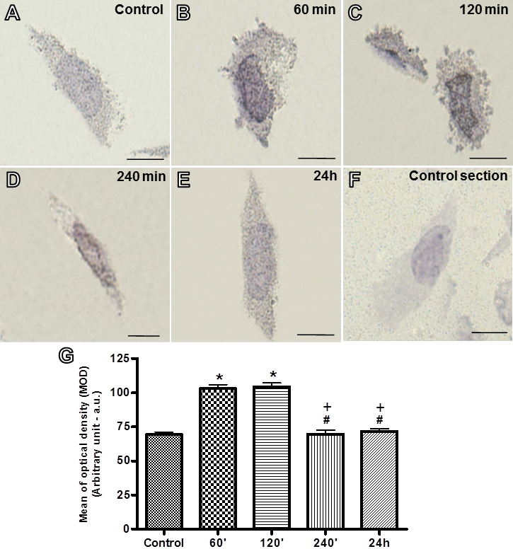
 Figure 5
of Mimura, Mol Vis 2012; 18:1583-1593.
Figure 5
of Mimura, Mol Vis 2012; 18:1583-1593.