Figure 1. Histopathological analysis of ocular tissues in T. gondii infection. Control group (A-C). Experimental group (D-I). Disruption of normal retina architecture, formation of gaps (*; B, C, E, and H), retinal detachment (hollow curved arrow; A, E, and F) and the presence of inflammatory cells (arrows) in control and experimental groups after 24, 48, and 72 h. Mast cell (filled
curved arrow) in sclera region (G). Blood vessel (v). Stain: Toluidine blue. Scale bars: 50 μm (A-F), 10 μm (G-I).
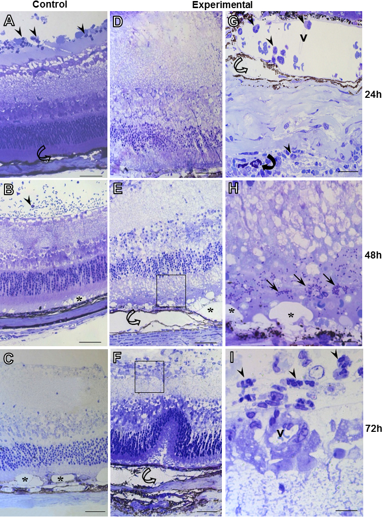
 Figure 1 of
Mimura, Mol Vis 2012; 18:1583-1593.
Figure 1 of
Mimura, Mol Vis 2012; 18:1583-1593.