Figure 6. Effect of tear collection method on protein profiles. A: Gel view of tear profiles collected from CT1, CT3, CT4, and CT6 by standard capillary technique without addition of physiologic
serum (LE: S-, left eye), and by the eye-flush technique involving the addition of physiologic serum (RE: S+, right eye).
B: PCA representation of tear protein profiles obtained from the same individuals. Left eye tears were collected by standard
capillary technique (light spheres) and right eye (dark spheres) by the addition of physiologic serum.
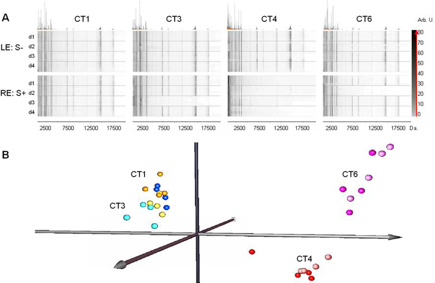
 Figure 6 of
González, Mol Vis 2012; 18:1572-1582.
Figure 6 of
González, Mol Vis 2012; 18:1572-1582.