Figure 1. Novel changes identified in myocilin in POAG patients. All the changes were identified in the heterozygous condition. The
mutated base is indicated with an arrowhead in the chromatograms. A: The chromatogram on the left demonstrates location of a synonymous variant (c.372 G>C, Leu124Leu) and a deletion mutation
(c.375, del G, R125SfsX158). On the right, the cartoons show all the known domains of normal MYOC and the truncated protein
resulting from the deletion, including aberrant 33 amino acids at the COOH-terminal end. B: The chromatogram on the left demonstrates location of a deletion mutation (c.819, del C, D273DfsX344). On the right, the
cartoons demonstrate the known domains of normal MYOC and the truncated protein resulting from the deletion, including aberrant
71 amino acids at the COOH-terminal end. C: The chromatogram on the left demonstrates location of a nonsynonymous variant (c.992 C>T, Ser331Leu). On the right, conservation
status of the residue (indicated by arrowhead) is shown in homologous protein in other species.
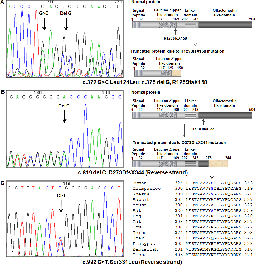
 Figure 1 of
Banerjee, Mol Vis 2012; 18:1548-1557.
Figure 1 of
Banerjee, Mol Vis 2012; 18:1548-1557.