Figure 7. Perlecan localization is altered in P2X7−/− stromas. Frozen corneas were sectioned and stained with antibody against perlecan, FITC-conjugated IgG, and To-Pro 3AM. Negative
controls were incubated with non-immune IgG instead of primary antibody, and To-Pro 3AM. Images are representative of three
independent experiments. A: Perlecan expression is increased throughout the stroma in P2X7−/− corneas. Inset: enlarged regions from the central stroma with enhanced signal to show detail of localization. Scale bar:
10 μm. B: Perlecan is localized to the basement membrane in WT corneas (arrow) but not in P2X7−/− corneas. Scale bar: 500 μm.
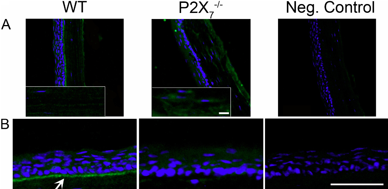
 Figure 7 of
Mankus, Mol Vis 2012; 18:128-138.
Figure 7 of
Mankus, Mol Vis 2012; 18:128-138.