Figure 6. Sulfation of glycosaminoglycans in WT and P2X7−/− corneal stromas. Collagen fibril length was measured, and the number of proteoglycan (PG) units along that length was counted
for each non-overlapping region (Anterior, Middle, Posterior) and summed for the entire cornea (Overall). Results were normalized
to number of PG units per 100 nm collagen for A: Undigested corneas, B: Corneas digested with Keratanase I, C: Corneas digested with Chondroitinase ABC, and D: Corneas digested with both Keratanase I and Chondroitinase ABC. A minimum of 75 measurements were performed for each region
of each group and digestion condition, and results were averaged and presented as ±SEM ** p<0.0001 and * p<0.05, one-way ANOVA
followed by Tukey’s post-hoc test.
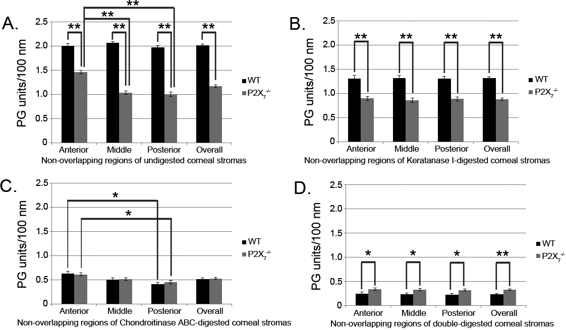
 Figure 6 of
Mankus, Mol Vis 2012; 18:128-138.
Figure 6 of
Mankus, Mol Vis 2012; 18:128-138.