Figure 3. KSPG localization is
altered in P2X7−/− stromas. Frozen
corneas were sectioned and stained with antibodies specific for
keratocan and lumican, followed by FITC-conjugated IgG (green)
and counterstained with To-Pro 3AM for nuclei (blue). Negative
controls were incubated with secondary antibody only and
counterstained with To-Pro 3AM. Images are representative of
three independent experiments. Keratocan (inset): enlarged
regions from central stroma with enhanced signal to show detail
of localization. Scale bar: 10 μm.
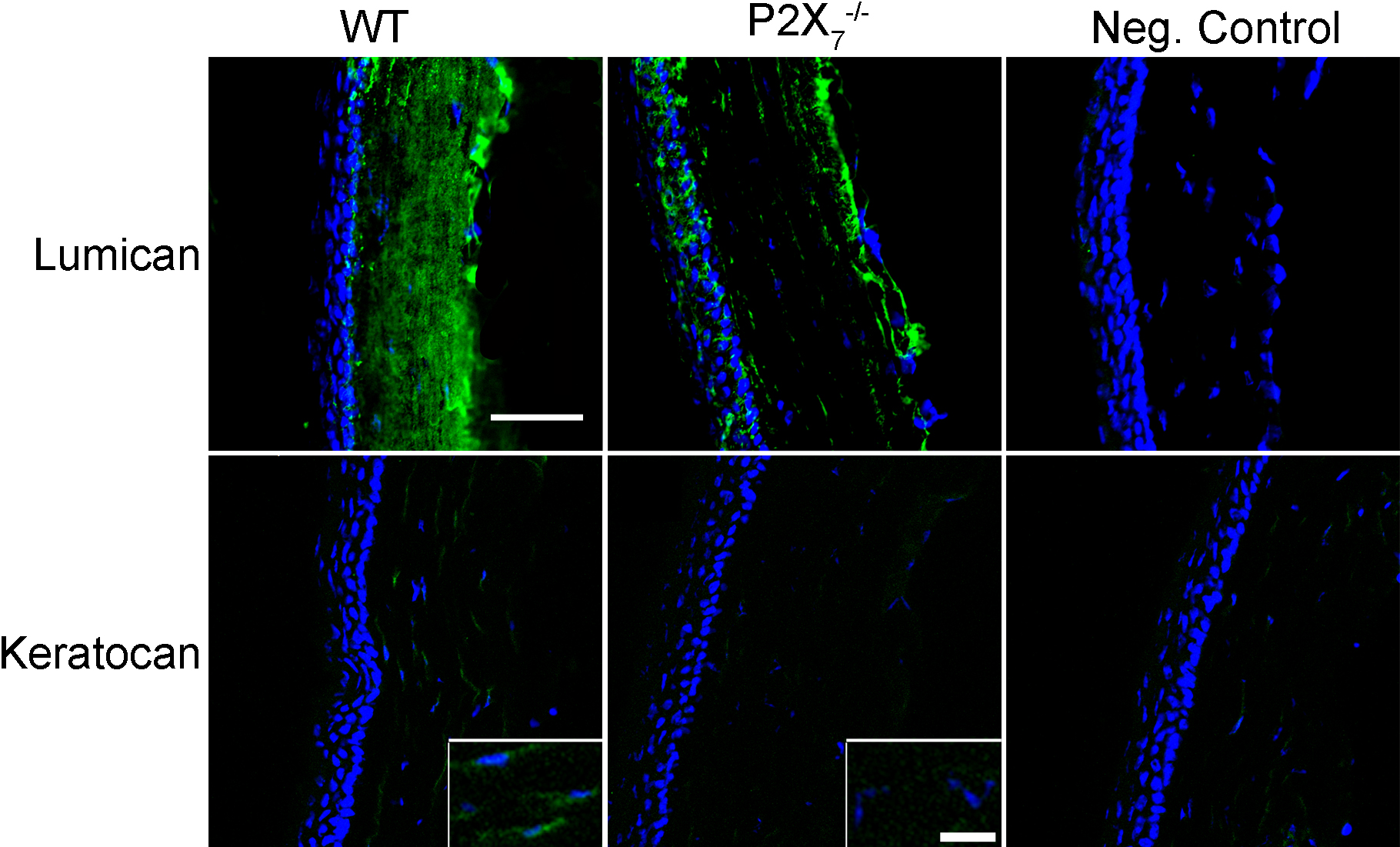
 Figure 3
of Mankus, Mol Vis 2012; 18:128-138.
Figure 3
of Mankus, Mol Vis 2012; 18:128-138.