Figure 6. Time course of 9-cis-retinal
treatment in Rpe65−/− mice on photoreceptor
specific proteins and the inhibitory effect of a protease
inhibitor mixture on M-opsin degradation in Rpe65−/−
mice and wild-type mice at three weeks in retinal explants. A:
The M-opsin, S-opsin, GNAT2, GNAT1, and rhodopsin levels treated
with 0.5 nM 9-cis-retinal for 6, 12, or 24 h are shown
with immunoblot in Rpe65−/− mice. B:
Quantitative analysis of effect of 9-cis-retinal on the
M-opsin protein level. Histograms indicate mean±SD (n=6),
**p<0.01. Western blots were performed to analyze the
expression of M-opsin protein in the retina-RPE choroids treated
with a protease inhibitor mixture (20 μM pepstatin A, 30 μM
E64d, and 10μM MG-132) for 12 h in Rpe65−/−
mice (C) and wild-type mice (D).
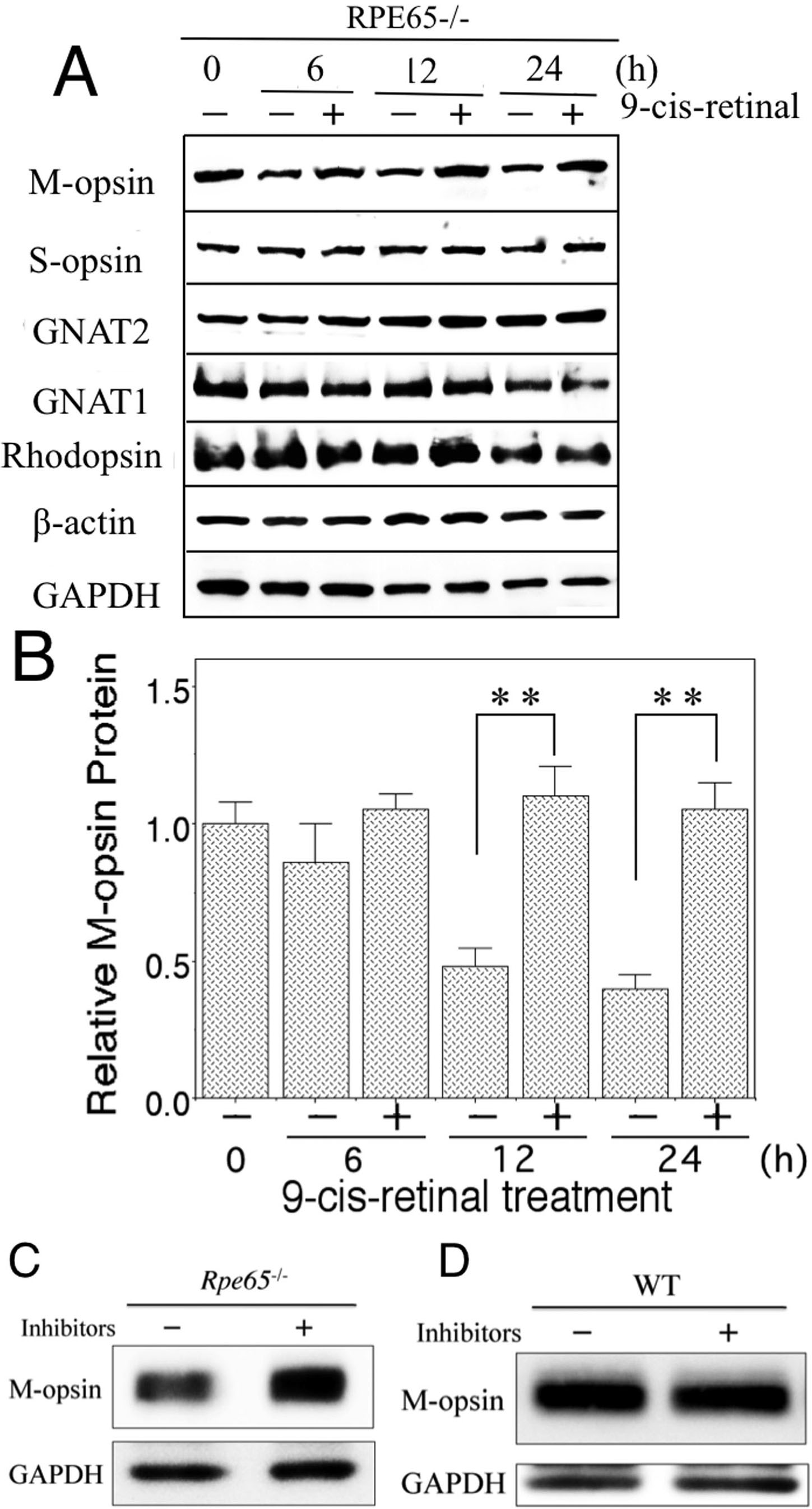
 Figure 6
of Sato, Mol Vis 2012; 18:1516-1525.
Figure 6
of Sato, Mol Vis 2012; 18:1516-1525.