Figure 5. Effect of 9-cis-retinal
on M-opsin expression in Rpe65−/− mice in
retinal explants. A: Retina-RPE choroids incubated with
0.5 nM or 5 nM 9-cis-retinal for 24 h were used for
western blotting to detect M-opsin protein. B:
Immunolocalization of M-opsin treated 0.5 nM 9-cis-retinal
for 24 h in the Rpe65−/− mice retina.
Immunoreactivity of M-opsin (red) was higher with 0.5 nM 9-cis-retinal
treatment compared to vehicle treatment in the cone outer
segments (arrows), ONL, and OPL (arrowheads) in the retina. C:
Histological findings of the density on the reactions labeled
anti-M-opsin antibody in a 400-μm-wide section of the retina.
Values represented mean ± SD (n=6). ***p<0.001. RPE, retinal
pigment epithelium; OS, outer segment; ONL, outer nuclear layer;
OPL, outer plexiform layer. Scale bars, 50 μm.
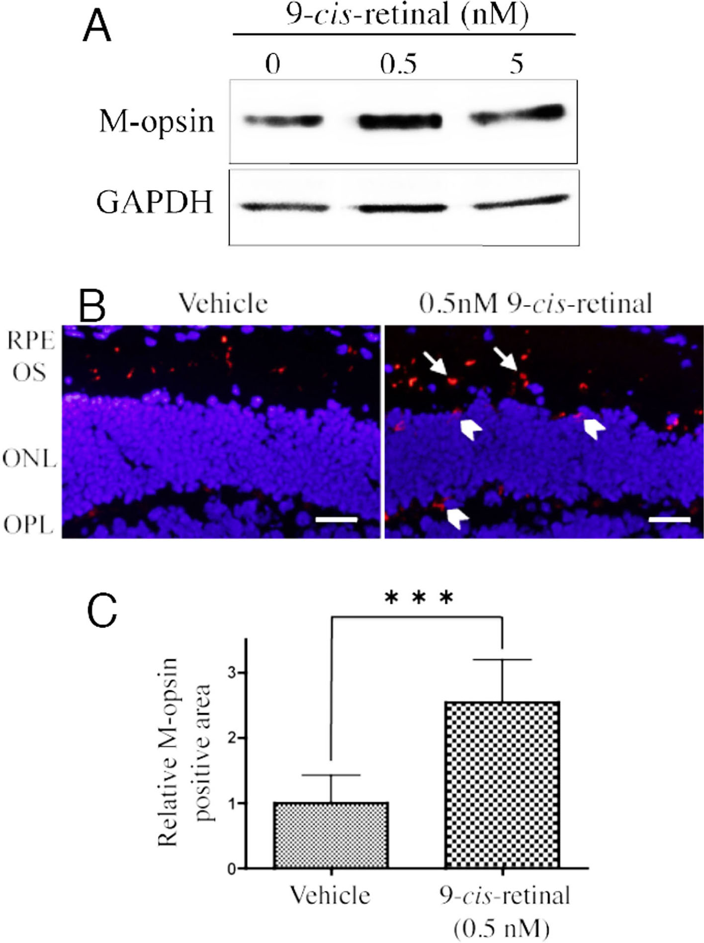
 Figure 5
of Sato, Mol Vis 2012; 18:1516-1525.
Figure 5
of Sato, Mol Vis 2012; 18:1516-1525.