Figure 1. Expression of SCF and c-kit in mouse cornea. A: Expression of the mRNAs of SCF and c-kit in mouse cornea. Total mRNA was extracted from cornea and brain tissues of WBB6F1-+/+mice. The mRNAs of SCF and c-kit were detected in corneal tissue with the predicted size of approximately 170 and 160 base pairs, respectively. The brain
was used as positive control (P). Negative control was carried with no DNA template (N). C:Cornea. B, C: Immunohistochemical detection of SCF and c-kit in mouse cornea. Corneal tissues from WBB6F1-+/+ were stained with anti-mouse SCF rabbit IgG (B) or anti-mouse c-kit rabbit IgG (C). In the cornea, a staining for SCF and c-kit was observed throughout the epithelium. Bar: 50 µm.
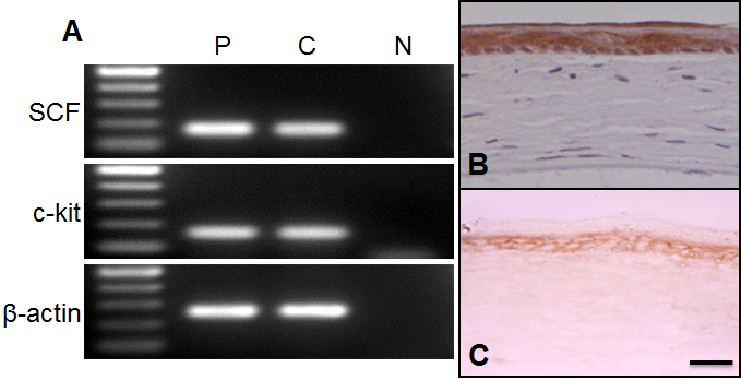
 Figure 1 of
Miyamoto, Mol Vis 2012; 18:1505-1515.
Figure 1 of
Miyamoto, Mol Vis 2012; 18:1505-1515.