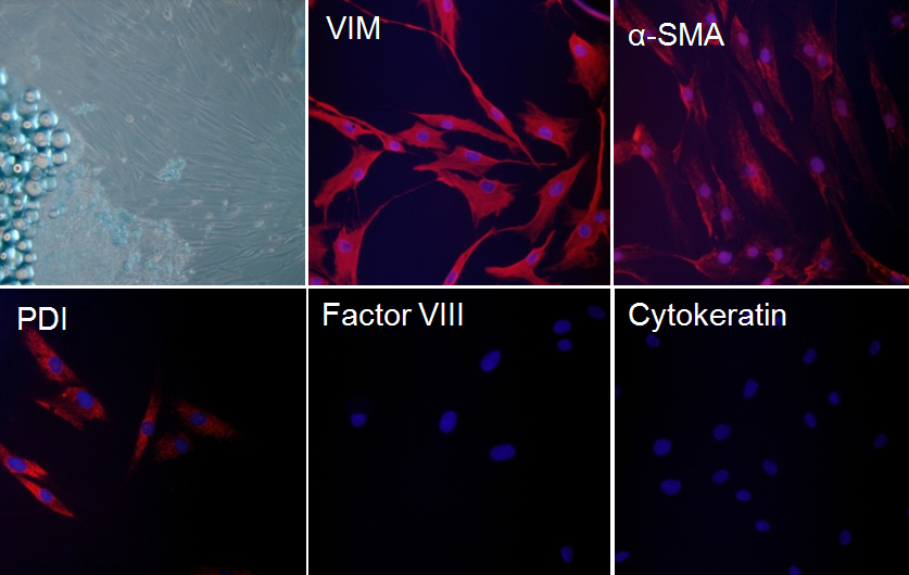
Figure 1. Characterization of orbital fibroblasts. Orbital fibroblasts were derived from retrobulbar fat tissue of patients with thyroid-associated
ophthalmopathy (n=5,
Table 1). Morphology was examined by phase-contrast microscopy and the expression patterns of fibroblast and non-fibroblast markers
were examined by immunostaining with antibodies against vimentin (VIM), α-smooth muscle actin (α-SMA), protein disulfide-isomerase
(PDI), factor VIII, and cytokeratin, respectively. Images were obtained from orbital fibroblasts of a representative patient
with TAO. Similar results were observed in orbital fibroblasts from all five patients with TAO.
 Figure 1 of
Paik, Mol Vis 2012; 18:1467-1477.
Figure 1 of
Paik, Mol Vis 2012; 18:1467-1477.