Figure 8. Established hypertension
causes and diabetes exacerbates retinal microvascular
degeneration. A: Representative images for retinal
trypsin digests stained with periodic acid-Schiff and
hematoxylin (PASH) to assess the development of acellular
capillaries in the established stage of spontaneous hypertensive
rats (SHR) and diabetic spontaneous hypertensive rats (SHR+D)
compared to control wistar group (W). Acellular capillaries were
defined as capillary-sized blood vessel tubes having no nuclei
anywhere along their length (arrows). B: Statistical
analysis for the average number of acellular capillaries per
group, showed higher numbers in the SHR and SHR+D groups by 2.8
and 5.6 folds, respectively, when compared to control W group
(n=4–5; *p<0.05). The SHR+D group was higher than the SHR
group by twofolds (#p<0.05). The SHR+D group was higher than
the SHR group by twofold (#p<0.05).
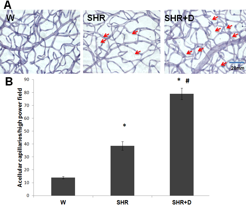
 Figure 8
of Mohamed, Mol Vis 2012; 18:1457-1466.
Figure 8
of Mohamed, Mol Vis 2012; 18:1457-1466.