Figure 6. Established hypertension
alone or in combination with diabetes stimulates retinal
inflammation. A: Representative image for western blot
analysis of retinal nuclear factor kappaB p65 (NFkB p65) protein
expression in the established stage of spontaneous hypertensive
rats (SHR) and diabetic spontaneous hypertensive rats (SHR+D)
compared to control wistar group (W). B: Representative
image for western blot analysis of retinal tumor necrosis factor
aplha (TNF-α) protein expression in the established stage of SHR
and SHR+D compared to control wistar group (W; n=4, *p<0.05).
C: Representative image shows results of western blot
analysis of retinal TNF-α protein expression. D:
Statistical analysis showed that TNF-α protein expression was
higher in the SHR and SHR+D groups by 3.4 and 5.4 fold,
respectively, relative to the control W group (n=4, *p<0.05).
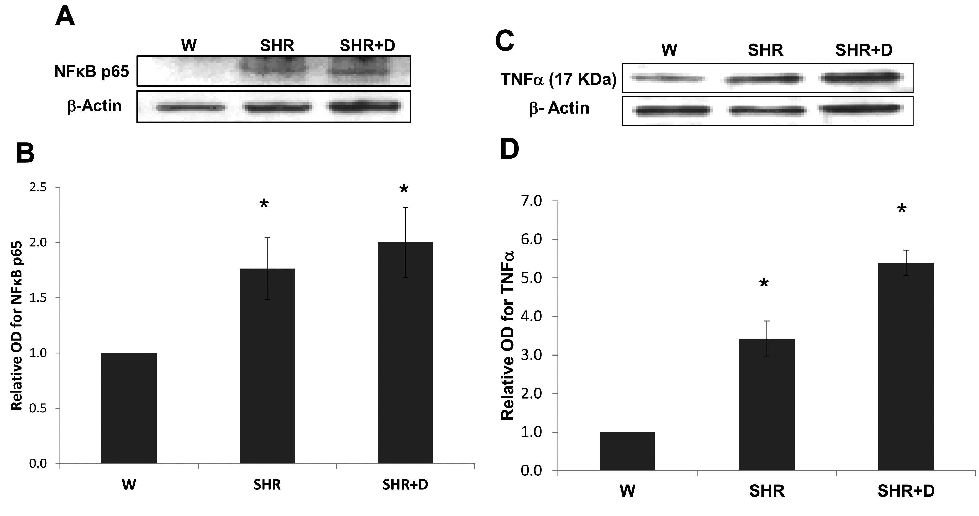
 Figure 6
of Mohamed, Mol Vis 2012; 18:1457-1466.
Figure 6
of Mohamed, Mol Vis 2012; 18:1457-1466.