Figure 4. Electropherograms of RAX
mutations. Asterisks denotes mutated bases. A:
Electropherogram of patient IV:1 (family A) showing a homozygous
c. 543+3A>G RAX mutation. B: Electropherogram
of patient IV:1’s father (family A) showing the heterozygous
mutation.
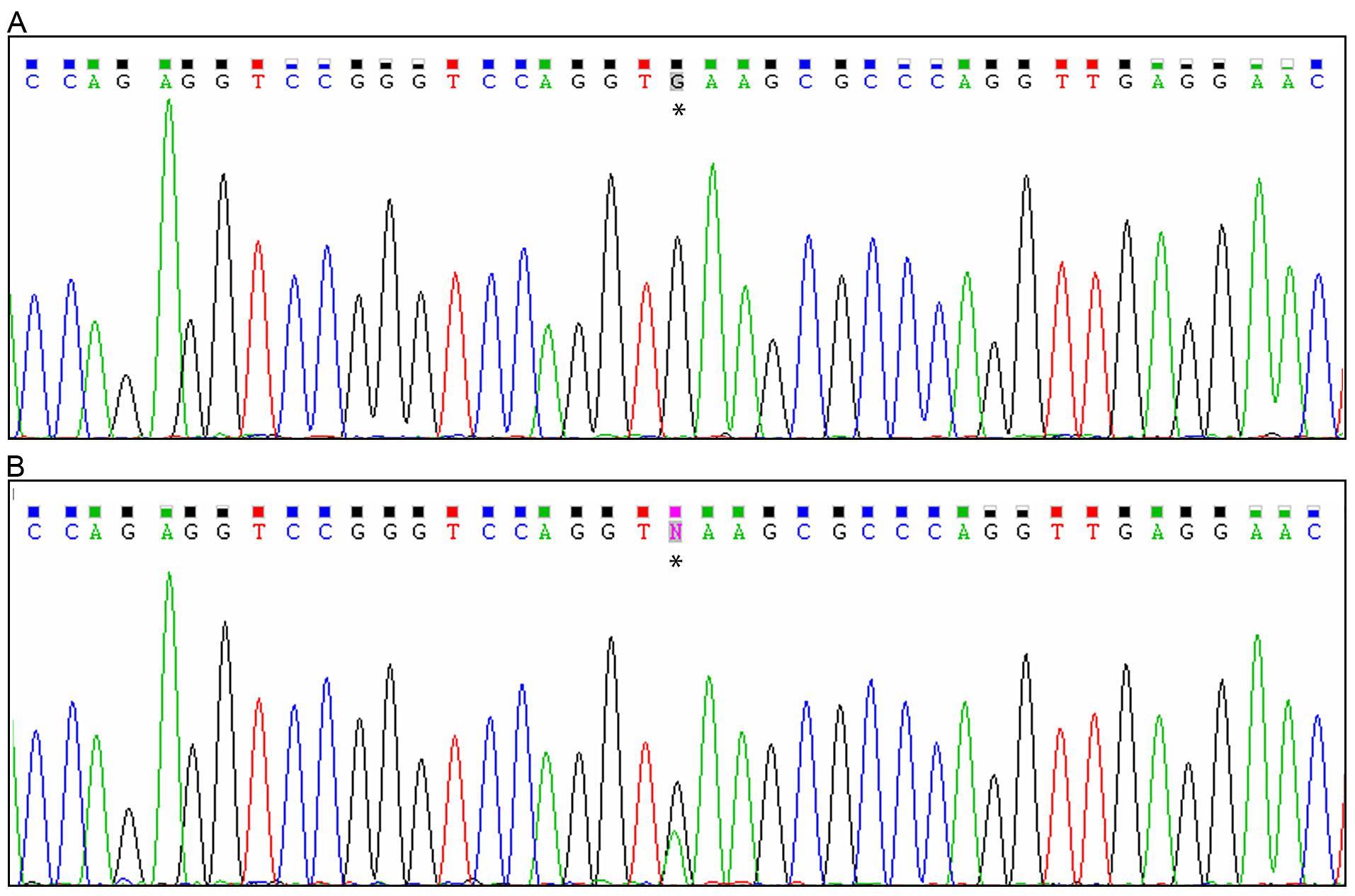
 Figure 4
of Abouzeid, Mol Vis 2012; 18:1449-1456.
Figure 4
of Abouzeid, Mol Vis 2012; 18:1449-1456.