Figure 3. Cerebral and orbital MRI
images. A: Family A, Patient IV:5. B: Family B,
affected daughter. Orbital hypoplasia. Coronal images,
T2-weighted. Agenesis of the eye globes. Rudimentary extraocular
muscles. Present and normal lacrimal glands. C: Axial
image. T1-weighted. Family A, patient IV:5. Absent frontal
sinus. D: Axial image. T2-weighted. Family B, affected
daughter. Corticosubcortical atrophy, predominantly in the
frontotemporal lobes, with ex vacuo dilation of the ventricles.
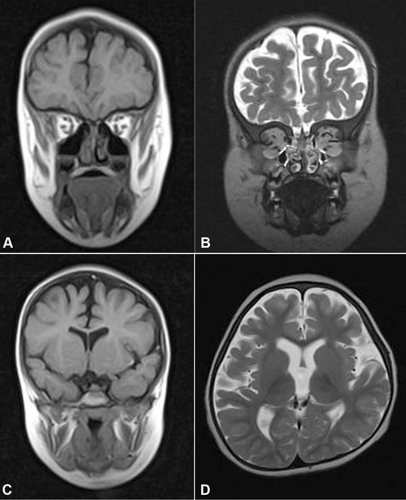
 Figure 3
of Abouzeid, Mol Vis 2012; 18:1449-1456.
Figure 3
of Abouzeid, Mol Vis 2012; 18:1449-1456.