Figure 3. Expression levels of FRMD7 at the indicated time points in Neuro-2a cells were confirmed by western blot. FRMD7 was monitored
by western blot in transfected Neuro-2a cells at the indicated time points. Samples (cells) from cultured Neuro-2a cells transfected
with pcDNA3.1 (+)-FRMD7-flag at different time points (24 h, 48 h, 72 h, and 7 days) were detected. The blots were probed
with the labeled antibody anti-flag. β-actin was used as an internal control.
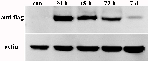
 Figure 3 of
Pu, Mol Vis 2012; 18:1428-1435.
Figure 3 of
Pu, Mol Vis 2012; 18:1428-1435.