Figure 3. TAZ protein clusters at the nucleus of proliferating epithelial cells. Conjunctiva epithelial cells were labeled for BrdU
(red) and TAZ (green), DAPI was applied as nucleus counter staining dye (blue). White arrows indicate a BrdU positive epithelial
cell also stained by high intensity of TAZ protein. Scale bar is 20 µm.
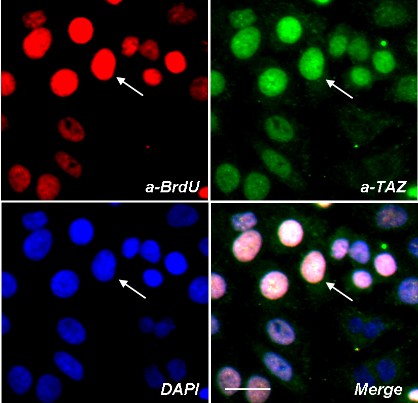
 Figure 3 of
Tan, Mol Vis 2012; 18:1402-1410.
Figure 3 of
Tan, Mol Vis 2012; 18:1402-1410.