Figure 3 of
Zhou, Mol Vis 2012; 18:1379-1383.
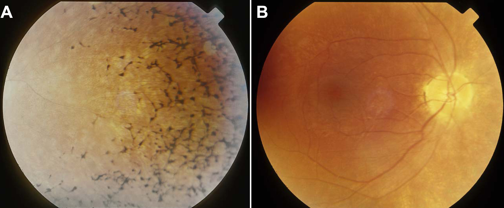
Figure 3.
Fundus photographs, OD, of patient II:2 at the age of 10.
A
: Bone spicules in nasal periphery.
B
: Pale optic nerve, vessel attenuation, and normal macula.
 Figure 3
of Zhou, Mol Vis 2012; 18:1379-1383.
Figure 3
of Zhou, Mol Vis 2012; 18:1379-1383.