Figure 5. This figure shows the phase contrast and fluorescent microscope images of Y79 cells grown on Poly(D,L-lactide-co-glycolide)
(PLGA) scaffold microparticles. A: Phase contrast microscope picture of Gelatin-coated PLGA microparticles under 10× magnification. B: Phase-contrast images showing cells attached to the microparticles (white arrows pointing Y79 cells attached to microparticles)
forming a three-dimensional growth over the microparticles under 10× magnification. C: Phase contrast microscopic image of 3-D growth of Y79 cells over scaffold microparticles (black arrow pointing to Y79 cells
attaché to microparticles) under 40× magnification. D: Fluorescent microscopic image showing the 3-D growth of Y79 cells (labeled with Celltracker dye) over scaffold microparticle
(white arrows pointing Y79 cells attached to microparticle) under 40× magnification.
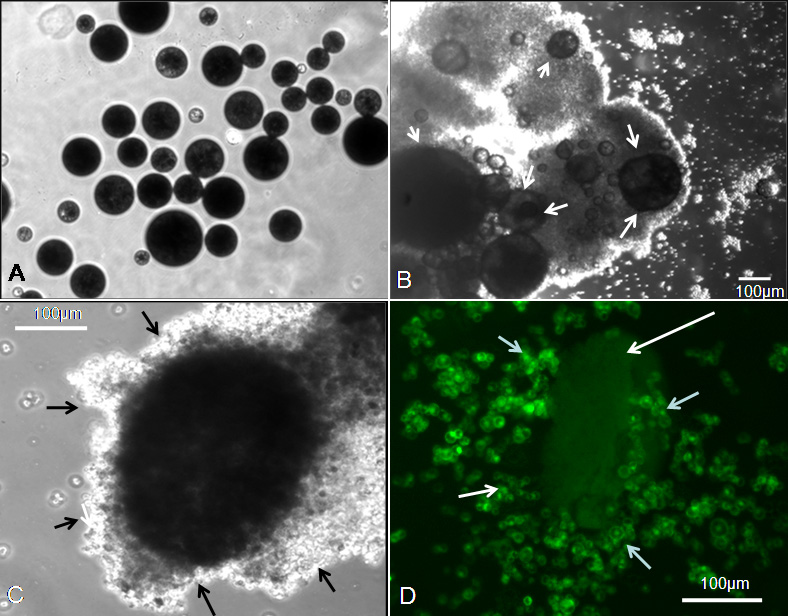
 Figure 5 of
Mitra, Mol Vis 2012; 18:1361-1378.
Figure 5 of
Mitra, Mol Vis 2012; 18:1361-1378.