Figure 2. This figure shows the scanning electron microscope picture of a Poly(D,L-lactide-co-glycolide) (PLGA) scaffold microparticle
containing 1.25% chitosan and 5% gelatin (100 mg PLGA, 1.25 mg chitosan, and 5 mg gelatin). Surface morphology of microparticles
was characterized by scanning electron microscopy (SEM). The average diameter of formulated microparticle in this study ranged
from 145 μm to 162 μm.
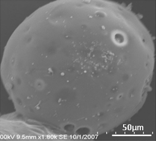
 Figure 2 of
Mitra, Mol Vis 2012; 18:1361-1378.
Figure 2 of
Mitra, Mol Vis 2012; 18:1361-1378.