Figure 3. The nucleus number beyond
retinal inner limiting membrane after HE staining. A:
The image of the retina in the negative control group under a
microscope. The endothelial cells were neatly arranged below the
inner limiting membrane. B: The image of the retina in
the positive control group under microscope. The endothelial
cells broke through the inner limiting membrane and grew in a
disorganized manner. C: The image of the retina in the
short hairpin RNA group under a microscope. The number of
endothelial cells increased, but only a minority of them broke
through the inner limiting membrane.
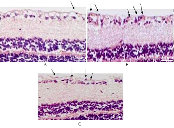
 Figure 3
of Li, Mol Vis 2012; 18:1354-1360.
Figure 3
of Li, Mol Vis 2012; 18:1354-1360.