Figure 2. Retina preparation by
fluorescein isothiocyanate-dextran heart perfusion. A:
The image of rat retinal angiography in the positive control
group under a fluorescent microscope. Tortuous and thick
abnormal new vessels were seen. B: The image of rat
retinal angiography in the positive control group under
fluorescent microscope. Fluorescent leakage of new vessel mass
was seen. C: The image of rat retinal angiography in the
gene interference group under a fluorescent microscope. Vessels
were distributed in a disorganized manner. D: The image
of rat retinal angiography in the gene interference group under
a fluorescent microscope. Tortuous and thick new vessels were
found. E: The image of rat retinal angiography in the
gene interference group under a fluorescent microscope. Two
layers of retinal vessels in normal distribution. F: The
image of rat retinal angiography in the gene interference group
under a fluorescent microscope. Deep vessels were normally
arranged as a net.
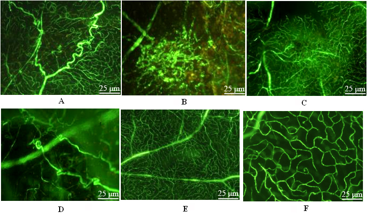
 Figure 2
of Li, Mol Vis 2012; 18:1354-1360.
Figure 2
of Li, Mol Vis 2012; 18:1354-1360.