Figure 1. Cell transfection detected
by fluorescence microscope. A: The image of the frozen
section of rat eyeball wall in the control group under a
microscope. Moderate green fluorescence was observed in the
retinal region close to the vitrea bulbus 4 days after blank
vector transfection. B: The image under a fluorescent
microscope. Green fluorescence was seen in the retinal region. C:
The image of the frozen section of rat eyeball wall in the gene
interference group under a microscope. Moderate green
fluorescence was seen in the retinal region close to the vitrea
bulbus 4 days after recombinant vector transfection. D:
The image under a fluorescent microscope. Green fluorescence was
seen in the retinal region.
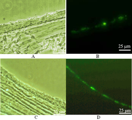
 Figure 1
of Li, Mol Vis 2012; 18:1354-1360.
Figure 1
of Li, Mol Vis 2012; 18:1354-1360.