Figure 3. Colocalization of
adrenomedullin (ADM) with cell specific markers was done to
accurately identify cell types with ADM-like immunoreactivity
(LI). In all figures, ADM-LI was localized near the
photoreceptor outer segments and in somata in the inner nuclear
layer (INL) and the ganglion cell layer (GCL). A:
Glutamine synthetase (GS, red) labeled Müller cells, and
colocalized with ADM (green) in their somata in the INL
(horizontal arrow). B: Protein kinase C α-like
immunoreactivity (PKCα-LI; green) was present in rod bipolar
cells and did not colocalize with ADM-LI (red). C:
Calbindin-LI (green) was localized in horizontal cells in the
INL and their process in the OPL, as well as in somata in the
INL and GCL. ADM-LI (red) colocalized with calbindin-LI in
somata in the lower tier of the INL that borders the IPL
(vertical arrow) and in somata in the GCL (diagonal arrow). D:
Calretinin-LI (green) was localized in amacrine cell somata in
the INL, in processes in the inner plexiform layer (IPL), and
within somata in the GCL. ADM-LI (red) did not colocalize with
calretinin-LI. E: neuronal nitric oxide synthase
(nNOS)-LI (green) was localized in select amacrine cell somata
in the INL, in processes in the IPL, and in somata in the GCL.
Some somata with nNOS-LI in the GCL colocalized with ADM-LI
(red) (arrow). F: Glutamate-LI (red) was localized near
the photoreceptor outer segments, in the OPL, in somata in the
INL and the GCL, and as diffuse staining in the IPL. ADM-LI
(green) colocalized with glutamate near the outer segments
(arrow). G: Glycine-LI (red) was localized in the INL,
in the OPL and the IPL, and in somata in the INL and the GCL.
Glycine-LI was not colocalized with ADM-LI (green) in any
somata, but there was some potential colocalization in puncta in
the IPL (arrow). H: gamma-aminobutric acid (GABA)-LI
(red) was localized to somata in the ONL, processes in the OPL,
somata in the INL, diffusely in the IPL, and somata in the GCL.
ADM-LI (green) did not colocalize with GABA-LI. Scale bars=20
µm.
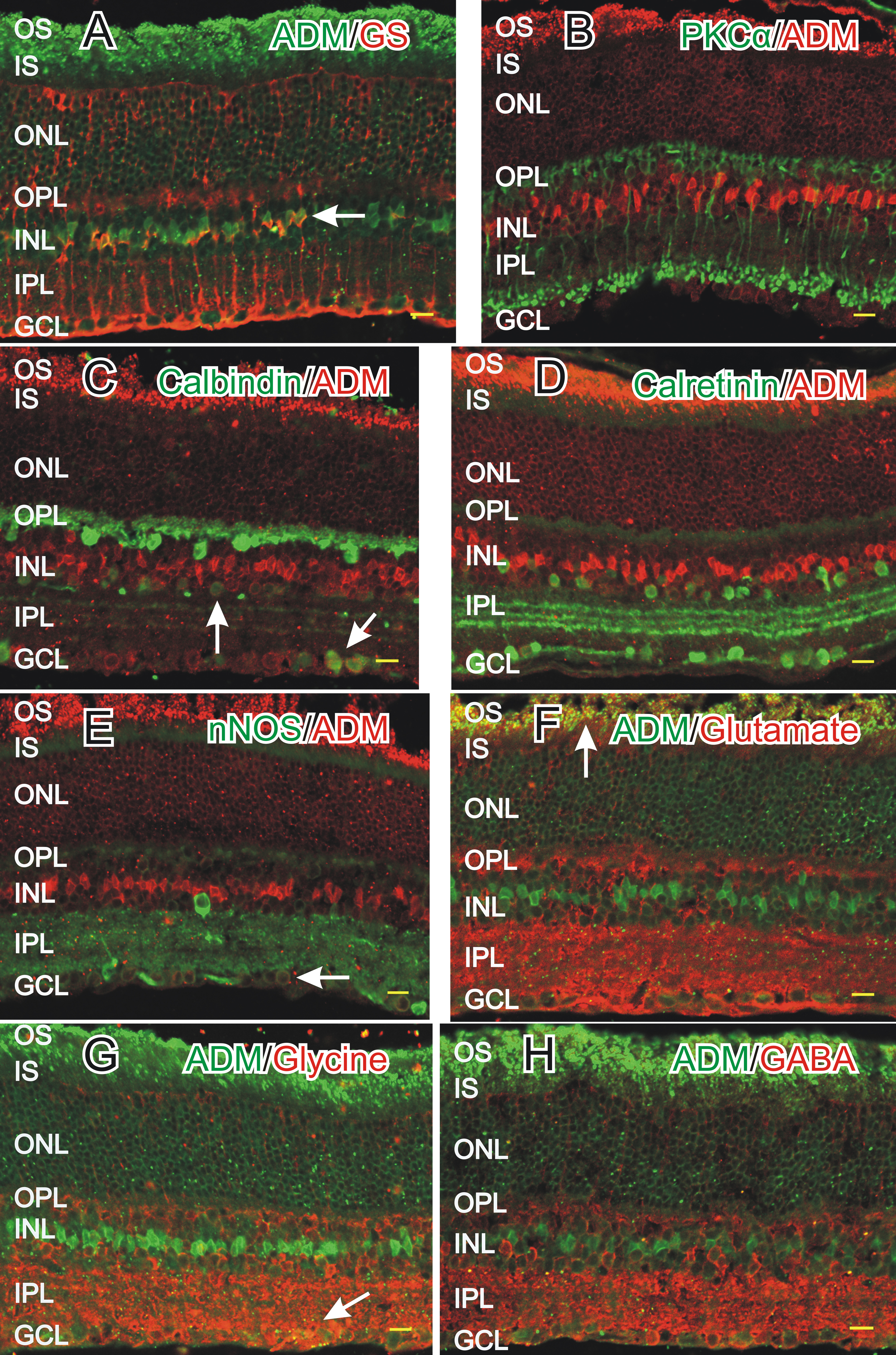
 Figure 3
of Blom, Mol Vis 2012; 18:1339-1353.
Figure 3
of Blom, Mol Vis 2012; 18:1339-1353.