Figure 2. Evaluation of cobalt
chloride induced cytotoxicity and the induction of hypoxia. A:
Hypoxia-induced growth arrest by cobalt chloride at various
concentrations (100–700 µM) in choroidal endothelial cells; the
concentration of cobalt chloride used for inducing hypoxia is
represented on the x-axis and the percentage of viable cells
normalized against control on the y-axis. B: The effects
of hypoxia induced by cobalt chloride on the cell cycle.
Choroidal endothelial cells (CECs) were treated with cobalt
chloride at various concentrations (0 µM, 100 µM, 200 µM) for 24
h and analyzed for DNA content; representative data are
displayed. (Mean+SE; n=3; *p<0.05).
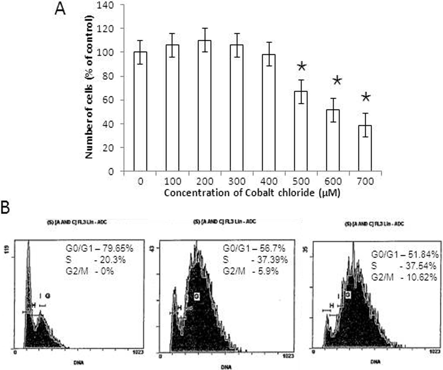
 Figure 2
of Balaiya, Mol Vis 2012; 18:114-120.
Figure 2
of Balaiya, Mol Vis 2012; 18:114-120.