Figure 3. Expression and
quantification of selected proteins in 661W cells treated with
caffeic acid phenethyl ester (CAPE) and H2O2.
A: Expression and quantification of heme oxygenase 1
(HO-1), cyclooxygenase 2 (COX-2), and IκBα proteins in 661W
cells was measured by western blot analysis. Proteins were
extracted and subjected to western blotting with anti-HO-1,
anti-COX-2, and anti-IκBα antibodies. Lane 1(NT): no treatment;
lanes 2 and 3 (caffeic acid phenethyl ester [CAPE]): CAPE
treated; lanes 4 and 5 (H2O2): H2O2
treated; lanes 6 and 7 (C+H): pretreated with CAPE, then with H2O2.
B: Quantification of COX-2 and IκBα in 661W cells with
western blotting. Quantification of COX-2 and IκBα was obtained
with densitometric analysis, and normalized with β-actin.
(n=3–6; *: p<0.05, by the Student t test).
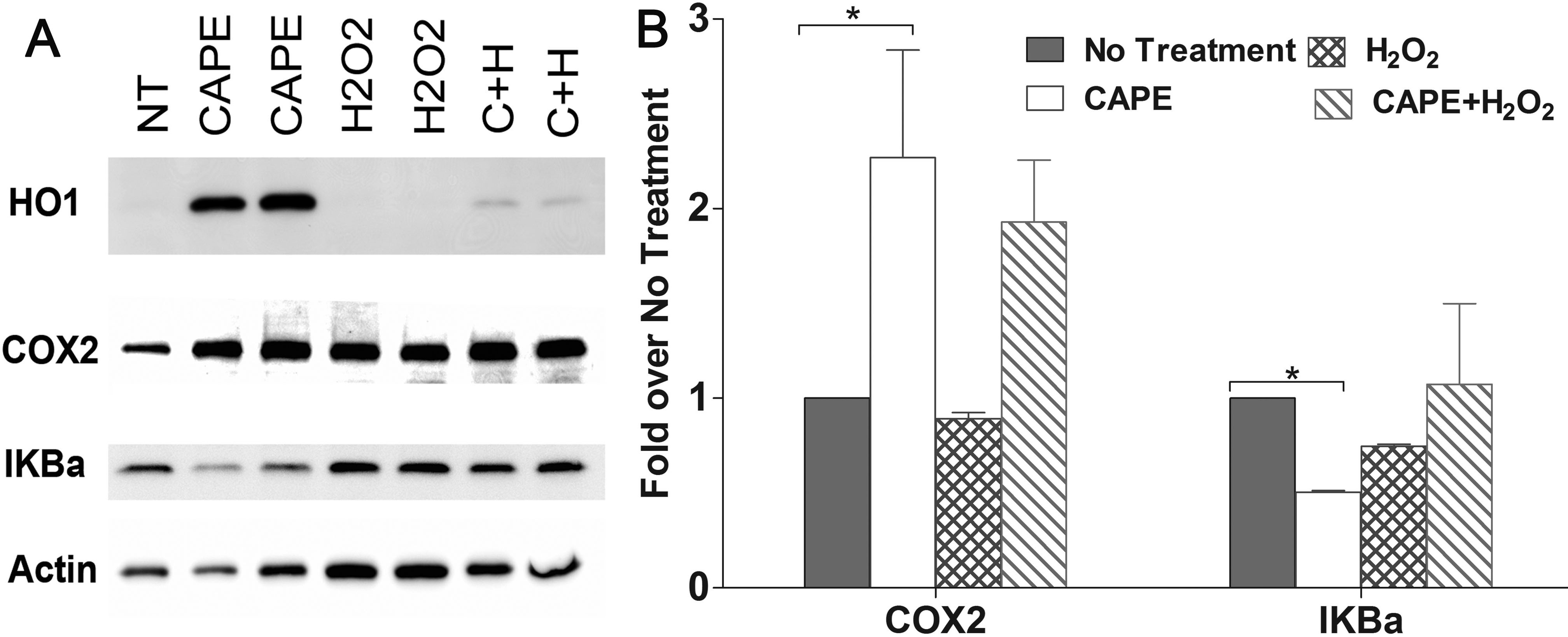
 Figure 3
of Chen, Mol Vis 2012; 18:1325-1338.
Figure 3
of Chen, Mol Vis 2012; 18:1325-1338.