Figure 4. Methylation percentages of cytosine-phosphate-guanine (CpG) sites in the collagen type Iα1 promoter region in scleras of monocular
deprivation and control eyes after four weeks of monocular deprivation. Numbers in parentheses on the x-axis are the locations
of the cytosine-phosphate-guanine (CpG) sites. Methylation percentages at sites 1, 3, 9, 14, 18, and 19 were significantly
greater in monocular deprivation–treated (MD-T) eyes than age-matched normal control (NC51) eyes. MD-T: monocular deprivation-treated eyes, MD-C: contralateral control eyes. *, p<0.05, **, p<0.01.
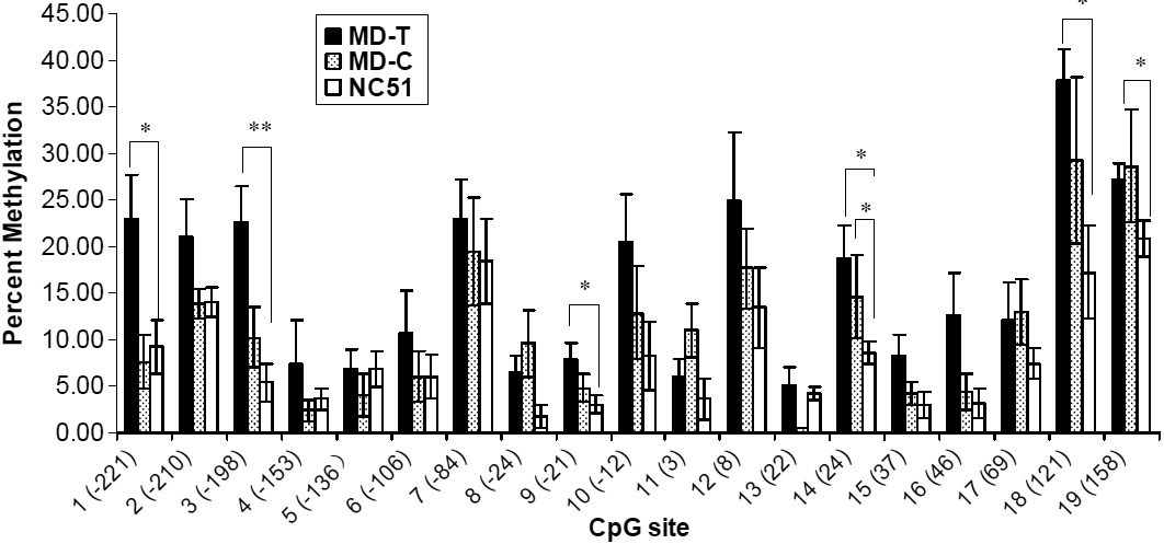
 Figure 4 of
Zhou, Mol Vis 2012; 18:1312-1324.
Figure 4 of
Zhou, Mol Vis 2012; 18:1312-1324.