Figure 4. Immunofluorescence staining of limbal stromal cells. The limbal stromal cells were positive for vimentin (200×; green), but
dim positive for SSEA-4 (100×; green). Expression for ABCG2 (100×), p63 (100×), Pax 6 (100×), AE5 (100×), CK 10 (200×), keratocan
sulfate (200×), α-SMA (200×), and Oct 3/4 (100×) proteins was found negative. The nuclei were counterstained either with propidium
iodide (red) or DAPI (blue).
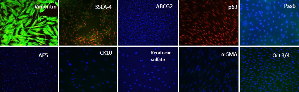
 Figure 4 of
Lim, Mol Vis 2012; 18:1289-1300.
Figure 4 of
Lim, Mol Vis 2012; 18:1289-1300.